Lab #6: Gel Electrophoresis and DNA Analysis- 2/21/19

Megan Hudson February 21, 2019
BIO 1106- 22
Lab #6: Gel Electrophoresis and DNA Analysis
Section I: Objective
The purpose of this lab was to perform gel electrophoresis and observe the DNA bands within the gel. Using these observations will help us conduct research on the soil ciliate biodiversity by analyzing the DNA sequence of the soil environment.
Section II: Procedure
- Start by practicing the technique of pipetting the 1 X loading buffer into the gel with Dr. Adair
- Be sure to have gloves on to prevent contamination with the Ethidium Bromide (since its a carcinogen)
- Place gel in the electrophoresis chamber in the correct orientation with the wells at the negative end of the chamber
- Pour 1X TAE running buffer over the gel to completely submerge the gel
- Assign wells to each DNA sample and DNA mass standard and draw a picture of what sample will go where.
- Pipette 9 ul of the DNA sample and 1 ul of the 10x loading buffer into the small green nano-centrifuge tube to make a 10 ul total 1x buffer
- Centrifuge the 10 ul total 1x buffer to fully mix the DNA with the buffer
- Pipette 10 ul of the purified DNA sample from Group 6 into Lane 1
- Pipette 10 ul of the purified DNA sample from Group 5 in Lane 7
- Load 5 ul DNA mass standard 1 (500 ng) in Lane 3
- Load 5 ul of the DNA mass standard 2 (15 ng ) in Lane 5
- Place top on the chamber and attach electrodes to the positive and negative ends
- Run the gel at 100 volts for approximately 20 minutes
- Take gel to the lab, place on the UV screen, and run the computer analysis program to visualize the DNA bands
- The ethidium bromide intercalated within the gel promoted the bands to fluoresce under UV light
- Using DNA mass standards, the bands of the DNA can be compared to the width and pigmentation of the standard to give an approximation of the mass of the fragments
- In the lab, we also performed absorbance tests over our purified DNA sample using the Nanodrop Spectrophotometer
- A 2 ul sterile water sample was placed on the optical surface to serve as a blank measurement
- A 2 ul drop of the purified DNA sample was placed on the optical surface
- The concentration and purity ratios were calculated
- The 260/280 ratio and the 260/230 ratio were analyzed
- The gel was returned to the refrigerator, and all used utensils were properly disposed of
- Work station was cleaned
Section III: Results and Observations
Gel Mock Up:
| Lane 1 | Lane 2 | Lane 3 | Lane 4 | Lane 5 | Lane 6 | Lane 7 | Lane 8 | Gel |
| Group 6 DNA | — | DNA Mas STD 1 |
—
|
DNA Mass STD 2 | — | Group 5 DNA | — |  |
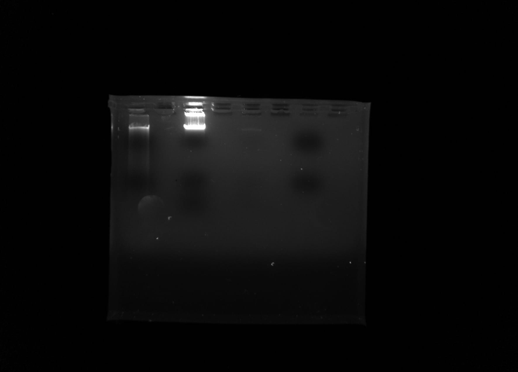
Gel under UV Light:
DNA Sample Nanodrop Spectrophotometer Analysis:
| Concentration of DNA based on Absorbance | 33 ng/ ul |
| 260/ 280 Ratio | 1.3 absorbance rate |
| 260/ 230 Ratio | 0.05 protein ratio |
Section IV: Where your sample was stored:
The agarose gel cast was stored in the refrigerator, and the remainder of the purified DNA sample was stored in the freezer at -20 degrees Celsius.
Section V: Conclusion and Future Steps
In this lab we performed gel electrophoresis by adding our purified DNA samples to the gel and using a battery to pull the negative charge of the DNA towards the positive end of the battery, creating bands of DNA based on the fragment size. When loading the 500 ng mass standard 1, 50 ul were pipetted instead of 5 ul, making it a 5000 ng solution and possibly skewing our comparison results. The Ethidium Bromide intercalated within the bands of the DNA, in a way mimicking a base pair. Placing the gel under a UV light allowed the bands of the DNA to fluoresce and allowed us to see the relative size and orientation of the DNA sequence. Unfortunately, our DNA sample did not produce any visible bands, therefore, our group has to perform DNA purification and run the gel again in open lab to hopefully produce better results. Since the smaller fragments will travel through the micro- inconsistencies to the bottom of the gel and the larger fragments will remain at the top of the gel, we should be able to observe the variation among the different soil sample’s DNA sequence. Since the goal of this lab is to develop research in soil ciliate biodiversity, observing and sequencing the DNA of the soil will allow us to better develop the community profile of the ciliates living within our soil samples.