March 8

Lab 8: PCR Results and Scientific Poster Design

Print Friendly, PDF & Email

Objective: 

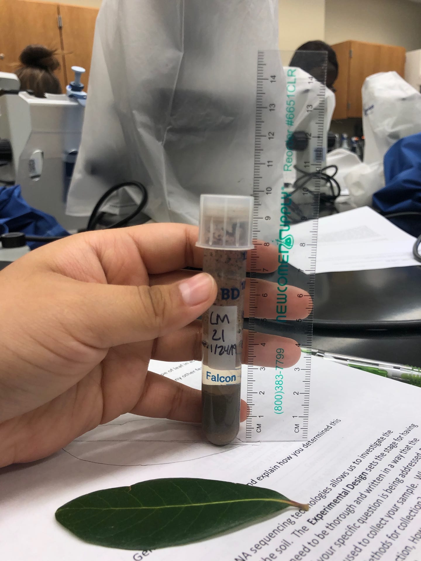
The main goal of this lab was to run our samples through gel electrophoresis in order to determine whether or not our samples qualified to be sent off to be sequenced. Furthermore, we took our midterm which allowed us to review everything we have covered so far in lab and make sure we remember it well. Lastly, we began drafting our scientific poster that we will present later in the semester.

Procedure:

  1. The 1.5% gel was previously prepared and already set up for us to begin pipetting
  2. Pipette 5 µl of 1 kb ladder into well #1 of the gel
  3. Pipette 10µl of your DNA sample into well #2
  4. Pipette 10µl of your control sample into well #3
  5. Then the other group’s control and DNA sample were pipetted into the next two wells
  6. Run the gel at 100V for 30 minutes
  7. Use the imaging software to view the results of gel

Data:

The first image shows our results from the imaging software. The second image shows our wells filled with our DNA and control groups.

Conclusion/Future Goals: 

The results show that our DNA was pure enough to show a high concentration on the imaging. One of the other two groups results also showed a high concentration of DNA. In the future, our DNA could be sent off to be sequenced. Furthermore, we are going to continue working on our scientific poster in order to have it ready to present when the time comes.

March 1

Lab 7: PCR amplification of DNA

Print Friendly, PDF & Email

Purpose: 

The main goal of lab today was to set up a PCR reaction using the eDNA that we collected from a Live Oak tree located on campus. Furthermore, we began drafting what we wanted our poster look like. This poster will contain all of the information that we have researched and data we have collected.

Procedure:

PCR Preparation

  1. we collected our tubes, one contained water, one contained v4 primer, and the other contained our DNA
  2. in a new tube, dilute the DNA by 10 by mixing 1μl of DNA with .9 μl of water
  3. collect two tubes containing 12.5μl of the master mix
  4. label one tube control and the other experimental (we labeled our TEST)
  5. make the solutions for each tube

control tube

6. add 1 μl of the v4 primer and 11.5 μl of water ( the contents of the tube should total to a total volume of 25 μl)

TEST tube

7.  add 1 μl of the diluted DNA, 1 μl of of the v4 primer, and 10.5 μl of water ( the contents of the tube should total to a total volume of 25 μl)

8. place the control and test tube in a rack and number the location of your tube on the worksheet provided

Poster 

For the poster, we essentially just drafted some potential ideas for what we wanted our poster to look like. It will include nearly all  the contents that a methods research paper would. We plan to add figures that will aid in describing our research and also pictures taken throughout the research process in lab.

Data: 

Our data showed that our DNA contained 598 ng/μl so we diluted it by 10 and calculated that we needed 1 μl of the diluted DNA which would contain 59.8 ng/μl.

Observations: 

When pipetting more than 10μl of a substance, we were advise to continue using the P10 pipette and cit the volume in half and pipette twice. This will help with accuracy and is more efficient than using a larger pipette which could lead to errors in the amount of volume transferred.

Conclusion/ Future Goals: 

This lab has allowed me to become more familiar with how to solve for different values using the C1V1=C2V2 formula. I have been able to understand the calculations more than I did when we first began using the formula. In the future, I plan to continue brainstorming on ideas for our poster that we will present. I will also continue to study for our in class quiz that we will have next week.

February 22

Lab 6: Gel electrophoresis and DNA Analysis

Print Friendly, PDF & Email

Objective:

The purpose of today’s lab was to determine if there was any DNA in our sample. We did this by performing gel electrophoresis and then further analyzing our data. We also used a spectrophotometer to test the absorbance of our DNA.

Procedure:

Gel electrophoresis:

  1. remove the comb from the gel
  2. remove black borders from the gel mold
  3. make sure your gel is placed in the buffer chamber in the correct orientation and covered with TAE buffer
  4. for each sample, in a microfuge tube, mix 9  μl of your DNA with 1 μl of loading buffer for a total of 10 μl
  5. mix the tube by tapping it or tapping it on the table.
  6. load 10 μl of your DNA sample into lane 8
  7. load 5 μl of DNA mass STD 1 in lane 3
  8. load 5 μl of DNA mass STD 2 in lane 4
  9. load any other samples in lane 2
  10. allow the other group to load their samples
  11. run the gel at 100V for ~20-30 minutes

Spectrophotometer:

  1. Clean the nano drop machine
  2. Add a blank ( we used water) onto of the small glass piece
  3. Run the spectrophotometer
  4. Clean off the water
  5. Add 2 μL of your pDNA to the piece.
  6. Run the spectrophotometer
  7. Collect and record the data

Data:

What was in each well?

The voltage was held at a constant 100V and the samples began to run at about 12 1/2 minutes.

Nanometer:

ng/uL- 598

A260/A280-1.42

A260/A230- 0.55

Observations: 

The data that we received was lower than what we were hoping for. For example, our A260/A280 was 1.42 but pure DNA should be about 1.8. Furthermore, our A260/A230 value was .55 but it should have been around 2. Some obstacles that we faced in this lab include pipetting our DNA into its well. We ended up performing 3 trials to pipette the DNA into the well in the gel but our last trial we performed with a DNA sample that was 2 μl DNA, 2 μl loading buffer, and 6 μl water. Our results showed a faint amount of DNA but we believe that if we had been able to test a larger amount of our DNA than it would appear much brighter on the gel imager.

Conclusion:

We were able to see DNA in our gel, although the results from the nanometer indicate that it was not completely pure. I really enjoyed this lab, it is really cool to see all of the technology that has been created to conduct research such as what we are doing now.

Future Goals:

In the future, I plan to continue to review key terms and protocols to prepare for the midterm. I also plan to add the procedures that were performed today to my methods.


February 15

Lab 5: DNA Extraction Part 2

Print Friendly, PDF & Email

Objective:

The goal of this lab was to perform the DNA purification procedure and make our Agarose gel. As we are beginning to finish up the experiment, we are now able to compile all of our notes from our lab notebook and use them to write our Methods.

Procedure:

DNA purification

  1. Add 1ml of the “crude DNA extraction” to a 15 ml tube, or other appropriate tube for mixing.
  2.  Add 2ml of warm DNA resin. Before adding it, mix the resin by inversion several times.
  3.  Set up a column bottom on a syringe barrel. Set the column and syringe barrel on the vacuum filtration manifold.
  4. Add half of DNA/resin to the column then turn on the vacuum. As soon as the liquid has been pulled through the column, add the remainder of your sample.
  5. Wash the column by adding 2ml 80% isopropanol and using the vacuum to pull it through. Begin by adding a small amount of isopropanol and allowing it to flow through before adding more. This may prevent the column from clogging. (repeat twice for a total of 6ml of isopropanol being added)
  6. Remove the column from the barrel and put the column in a clean 1.5ml tube.
  7. Spin at 8000 x g for 5 min to remove residual isopropanol. (balance the centrifuge!)
  8. Take the column out of the 1.5ml tubes and put it in the 80C+ heat block for 30s-1min. Do not exceed 1 minute.
  9. Put the column in a new 1.5ml tubes. Add 50 μl of diH2O to the column.
  10. Incubate 1min.
  11. Spin at 8000 x g for 1 min.
  12. Throw away the column and store the tube on the rack.
Agarose gel
  1. Add 4ml of 10x stock TAE and 36ml of deionized water in a centile vile.
  2. Add 0.4g of agar powder to a 125ml Erlenmeyer flask
  3. Add the TAE solution to the flask.
  4. Heat in the microwave until the solution is clear and small bubbles come off the bottom when swirled (about 1 minute)
  5. Add 2μl of ethidium bromide to the flask.
  6. Place solution in flask into gel cast then let it solidify, this should take approximately 30 minutes.
  7. Pour 1x TAE on top of the solidified gel until the gel cast is completely full.
  8. Place the gel in a plastic bag with a small amount of the buffer then refrigerate.

Data:

We calculated that in order to make a 1% Agarose gel, we would need 2ml of 1xTAE and .4g of Agarose.


I have included some pictures taken during the duration of lab. In the first picture I am holding the 15 ml tube containing the DNA/resin liquid. The second picture is an image of the flask containing the components used to make the Agarose gel. Finally, the last picture shows the vacuum apparatus that we used.

Conclusion: 

We spent about half of lab working on the math problems on the QTM. I think this is very important because we need to be able to calculate the different amounts of substances we need to make a desired solution. Additionally, we are now ready to continue our lab next week by using the Agarose gel we prepared to look at the DNA.

Future Goals:

I am excited for next lab. I am hoping that we will see DNA in our Agarose gel. Additionally, now that we are through most of our experiment I can begin writing my methods draft for our final report. I look forward to continuing to learn about the diversity of our soil.

February 8

Lab 4: DNA Extraction

Print Friendly, PDF & Email

Objective:

The goal of this lab was to determine the texture of our soil and identify the Genus species of our tree.  Additionally, we began the DNA extraction process for our soil samples.

Procedure:

Determining Soil Texture Cont.

  1. Use a ruler to measure the % sand, silt, and clay. This procedure works best by taking a clear picture of the tube and zooming in on the sedimentation lines.
  2. How can you determine the % of each type of soil particle?
  3. Use the USDA triangle and Soil Type Calculator to determine the texture of your soil.
  4. Record your soil type

Silica Bead DNA Extraction

  1. weigh 1 gram of soil and 1 gram of silica beads, transfer into an autoclaved mortar and pestle an grind finely for about 5 minutes
  2. add 2mL of the DNA extraction buffer and 10 mg of powdered activated charcoal to the soil-glass mixture and mix it by pipetting several times
  3. transfer the contents into a 2mL eppendorf tube
  4. incubate the tube in a hot plate at 65C for 10 minutes.
  5. centrifuge the tube at 12000g for 5 minutes
  6. extract as much of the supernatant as possible and transfer it into a fresh eppendorf tube

Out of our group of three, we decided to use Holli’s soil. In this procedure different components we’re added to aid in the extraction process. For example, the silica beads were added to help break down the soil. Furthermore, the charcoal was added to further remove impurities from the mixture.

Data: 

I determined that my soil was 32% sand, 64% silt, and 4 % clay. Therefore I would classify the texture of my soil as Silt Loam.

We classified our tree as a Quercus Virginiana Live Oak because of its evergreen leaves. We also used inaturalist and the other tree ID website to help confirm our classification.

Observations:

Last week during lab I was able to find ciliates in my non-flooded plate. I attempted to culture the ciliates but had no luck in finding any this week when I observed the well plates. Although, I was able to observe what we believe to be flagellates in my culture.

Conclusion: 

I am continuously learning about trial and error in lab. This week our lab group had to re-do the DNA extraction procedure because it did not work out for us the first time. Luckily, the second time around we knew what to do and how to do it better than the first time.

Future Goals:

In the future I am excited to continue the DNA Extraction process. I also plan to begin writing my methods draft in order to keep the events of lab clear and organized.

January 31

Lab 3: Ciliate Isolation

Print Friendly, PDF & Email

Objective:

The main goal of lab today was to find and observe ciliates in our non-flooded plates or test tubes then take pictures and videos and if possible, culture them.

Procedure:

% water content: 

  1. weigh empty Petri dish and record mass
  2. add “wet” soil to Petri dish and record mass
  3. determine weight of wet soil
  4. let soil sit for the allotted time 
  5. weigh the Petri dish with the soil and record mass
  6. determine % water content by using the formula wet soil-dry soil/wet soil x 100

Soil texture: 

Last week in lab we added about 3 mL of soil in a labeled 10 mL falcon tube and filled it to the 8mL mark with DI water. This week we proceeded to add a drop of dispersion agent and mixed it thoroughly using the vortex. Next week, when the soil has settled, we will determine the soil texture.

Ciliate discovery:

Take small samples from NFP or falcon tube using micropipette and observe using microscope. If ciliates are found, use micropipette to capture them and add to a 24 well plate to culture them.

Data:

The percent water content of my soil was 6.2%.

Observation: 

All members of my group said the ciliates that they’re seeing the most of are very small and peanut shaped, but occasionally I would see a bigger circle shaped ciliates swimming around on its own. I’m interested to know how these different ciliates interact or if they interact at all .

Conclusion/ Future Goals: 

Although I was not able to capture a great picture of the ciliates that I observed, I was able to culture my sample. I’m excited to check back next week and see if I was actually able to culture the ciliates that I found effectively. Also, I’m interested in knowing if percent water content and soil texture has an effect on how many ciliates we find or their behavior.


January 24

Lab 2: Metabarcoding and Scientific Presentations; Soil Collection

Print Friendly, PDF & Email

Purpose: 

The main goal of today’s lab was to present information that we collected over our articles. By presenting our information and listening to the information others collected we were able to learn more about the process of metabarcoding. Furthermore, another goal in lab was to set up our non-flooded plates with our soil samples, set up % water test , and observe the pH of the soil extract.

Procedure:

Soil pH

  1. place about 3 mL of soil in a labeled 10 mL falcon tube and fill to the 8mL mark with DI water
  2. mix thoroughly for 3-5 minutes
  3. remove about 1 mL of water from the top of the tube and transfer it to a microfuge tube
  4. spin the tube in the centrifuge for about 1 minute to to pellet the soil
  5. test the pH of the soil water using a pH strip and record result after comparing the color

Non-flooded plate 

  1. put 10-50g of soil in a Petri dish (just enough to cover the bottom of plate)
  2. saturate with distilled water
  3. observe your soil using the dissecting scope and record observations

Data:

pH- 6.0

Conclusion/ Future Steps:

I enjoyed learning various things from everyones presentations today. I think being able to become familiar with some of then concepts that we will be applying to our research will be useful for us. I wasn’t able to see anything in my non-flooded plate this lab, but I am hoping that next lab i’ll be able to extract some of the soil water and observe that. Additionally, in a future lab we will be calculating the % water of our soil samples.


January 17

Lab 1

Print Friendly, PDF & Email

Leslie Morales

1/17/19

Purpose:

The purpose of this lab was to get a brief overview of what we would be doing this semester in lab and how our research will aid us in answering our big question.  We also reviewed articles concerning the challenges in metabarcoding soil ciliates.

Summary:

Today in lab we reviewed a powerpoint which explained what we would be researching this semester. This semester we will be studying the biodiversity of ciliates by extracting eDNA using single isolate identification, metabarcoding, and metagenomics. Furthermore, we will be conducting research using soil from the trees here on campus. We also broke off into groups and were assigned an article to read. Our article was called Spatial genetic variation, phylogeography and barcoding of the peritrichous ciliate Carchesium polypinum. Our group then reviewed the article and created a powerpoint which presents the answers to some key questions about the article.

Goals for the Semester:

My goals for this semester are to learn more about soil ciliate biodiversity and the importance of soil ciliates. Furthermore, I hope to grow in my lab skills as well as continue to learn as we go through our research process. I am also very excited to be able to work with the trees here on campus and be able to potentially aid in providing useful information about the trees.

November 16

Lab 13: Ciliate Isolation and Characterization

Print Friendly, PDF & Email

Objective: 

The goal for lab today was to determine the soil type by calculating the percentages of the components that make up our soil samples. Furthermore, we continued to search for and identify ciliates.

Purpose:

This lab allowed us to determine what kind of soil our ciliates have come from and therefore we are able to see what conditions soil ciliates live in. additionally, we continued to utilize our lab techniques to try to isolate and observe ciliates.

Procedure:

Determining Soil Type 

  1. Retrieve soil tube sample
  2. Take a picture of the sample by a ruler
  3. Determine the total of soil and record data
  4. Determine the total amount of silt, clay, and sand then record data
  5. Calculate the amount of each silt, clay, and sand by dividing the amount of each different sedimentation by the total amount of soil
  6. Determine the soil type using the USDA triangle

Soil Ciliate Discovery 

  1. Obtain your petri dish.
  2. Place three drops of the soil sample water on the concavity slide
  3. Use the dissecting microscope to look for ciliates
  4. If the ciliates are moving to fast use methyl cellulose or if there’s too many soil, dilute with water. To view the ciliates better use iodine to dye the drop
  5. Take photos and videos of the ciliate and try to classify the discovered ciliate

Or

  1. Obtain your tray with your cultured sample
  2. Place three drops of the cultured sample on the concavity slide
  3. Use the dissecting microscope to look for ciliates
  4. If the ciliates are moving to fast use methyl cellulose or if there is too many soil, dilute with water. To view the ciliates better use iodine to dye the drop
  5. Take photos and videos of the ciliate and try to classify the discovered ciliate

Data:

My soil was composed of 42.31% of sand, 46.15% of silt, and 11.54% of clay. Therefore, my soil type was Silt Loam.

Observations:

The ciliates that I observed in my cultured sample contained several small, round ciliates. For the first part of lab I spent time observing drops from my cultured sample and for the latter half of lab I observed drops that I pipetted from my Petri dish. In drops from my Petri dish I was able to observe more types of ciliates rather than just the small, round ones.

Storage: 

We stored our cultured samples in a drawer at our lab table and returned microscopes to their appropriate places. Lastly, we returned our soil sample tubes back to the test tube rack and we rinsed the concavity slides and left them to dry.

Conclusion and Future Goals:

I was able to effectively culture my soil sample and use it to observe ciliates in an easier manner. I was also able to determine what type of soil my sample was by using our soil sample tubes. In the future I hope to take the information I’ve compiled over the past view weeks and use it to determine what kind of ciliates I have been observing. Furthermore, I plan to use my data in our group presentations next week.


November 9

Lab 12: Ciliate Classification

Print Friendly, PDF & Email

Objective: 

The main goal of this lab was to find ciliates and classify them into major classes using morphology. Furthermore, we began the process to determine the soil texture of our soil samples.

Purpose:

The purpose of this lab was to use our skills that we have developed over the semester to find a ciliate in our samples. Furthermore, we were allowed and encouraged to use methyl cellulose or a dye to see the ciliates better. Lastly, if we found ciliates we cultured them in hopes for them to grow.

Procedure:

Determining Soil Texture 

  1. remove any sticks, leaves, or other debris from your soil sample
  2. add soil to the 4ml mark in a falcon tube
  3. add water and mix vigorously
  4. add 1 drop of dispersing agent and re-mix using the vortex
  5. place the falcon tube in the rack until next lab

Soil Ciliate Discovery

  1. set up the dissection microscope
  2. obtain a clean slide
  3. pipette 3  drops of 10 μl of the excess water from the soil samples in the Petri dish onto the clean slide
  4. observe under microscope
  5. take videos and pictures if a ciliate is found

Observations:

IMG_7158-qz7s5g

Conclusion: 

I was able to find ciliates in my sample this lab! In a single drop I observed about 4 of the worm-like ciliates, 1 of the foot shaped ciliate, and several of the small round ciliates. Also, I was able to watch the worm-like ciliates eat. Furthermore, I was able to spend a good portion of the lab watching how the behavior of these three ciliates varied. Lastly, I was able to culture my ciliates.

Future Goals: 

In the future, I plan to stain a drop of my sample in attempt to see the ciliates better. I am hoping that my the ciliates in ym culture grow so that I am able to observe them next lab. Lastly, I am hoping that next lab I am able to spend a little more time trying to identify what type of ciliates I see.