Objectives:
1. To learn how to properly use a microscope, including:
-correctly handling the microscope when moving it from one location to another
-adjusting the focus and zoom knobs of the microscope
2. To be introduced to ciliates, as well as how to view and observe them, and to obtain basic information about different types of ciliates.
3. To gain experience conducting a lab, including:
-following procedures
-recording data
Purpose:
The purpose of the Ciliate Challenge lab was to observe 6 different types of ciliates, and to identify their names based on observations made along with identification sheets, which contained information about the distinct characteristics of different ciliates. To identify the ciliates species, one needed to observe the shape, color, size, and behavior of each ciliate they were observing. This lab involved developing the skill to use a microscope properly, as well as making detailed observations, and finding results based on those observations.
Procedure:
1. Set up the area in which the lab would take place. Make sure that hallways are clear of any objects. Ensure that the pipettes are clean, and that the wells being used are not contaminated.
2. Set up the microscope. Uncover the microscope and adjust the location of the device. Adjust the two lenses to be the same width as the space between the observers eyes. Plug in the microscope, and have the focus set to the lowest setting at the beginning.
3. Designate which unknown ciliates each member of the group will observe and attempt to identify.
4. Label the wells that will be used. Use the corresponding pipette to transfer the ciliate into the well. Be sure not to contaminate either the pipette, test tube, or well(s).
5. Use the zoom and focus knobs to obtain a crisp, close view of the ciliates in the well.
6. Record observations based on the guidelines: shape, relative size, movement, characteristics, sketch.
7. Use the information provided in the lab binder to attempt to identify each ciliate observed.
8. Collaborate and share data with each lab group member.
9. Reset the microscope arm to a better height if necessary, turn of the light, and unplug and cover the microscope.
10. Rinse out the wells with water, then spray bleach, and then place the well to dry.
11. Use the Windex to clean the table, then dry with paper towels.
Observations:
| Unknown # |
Shape |
Relative Size |
Movement |
Characteristics, Location |
Identification |
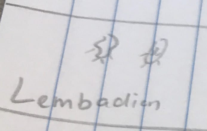
| 1 |
circular, round |
smaller than other ciliates |
very fast |
difficult to keep in view, very fast speed; located near surface |
unable to be identified due to speed and small size |
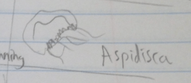
| 2 |
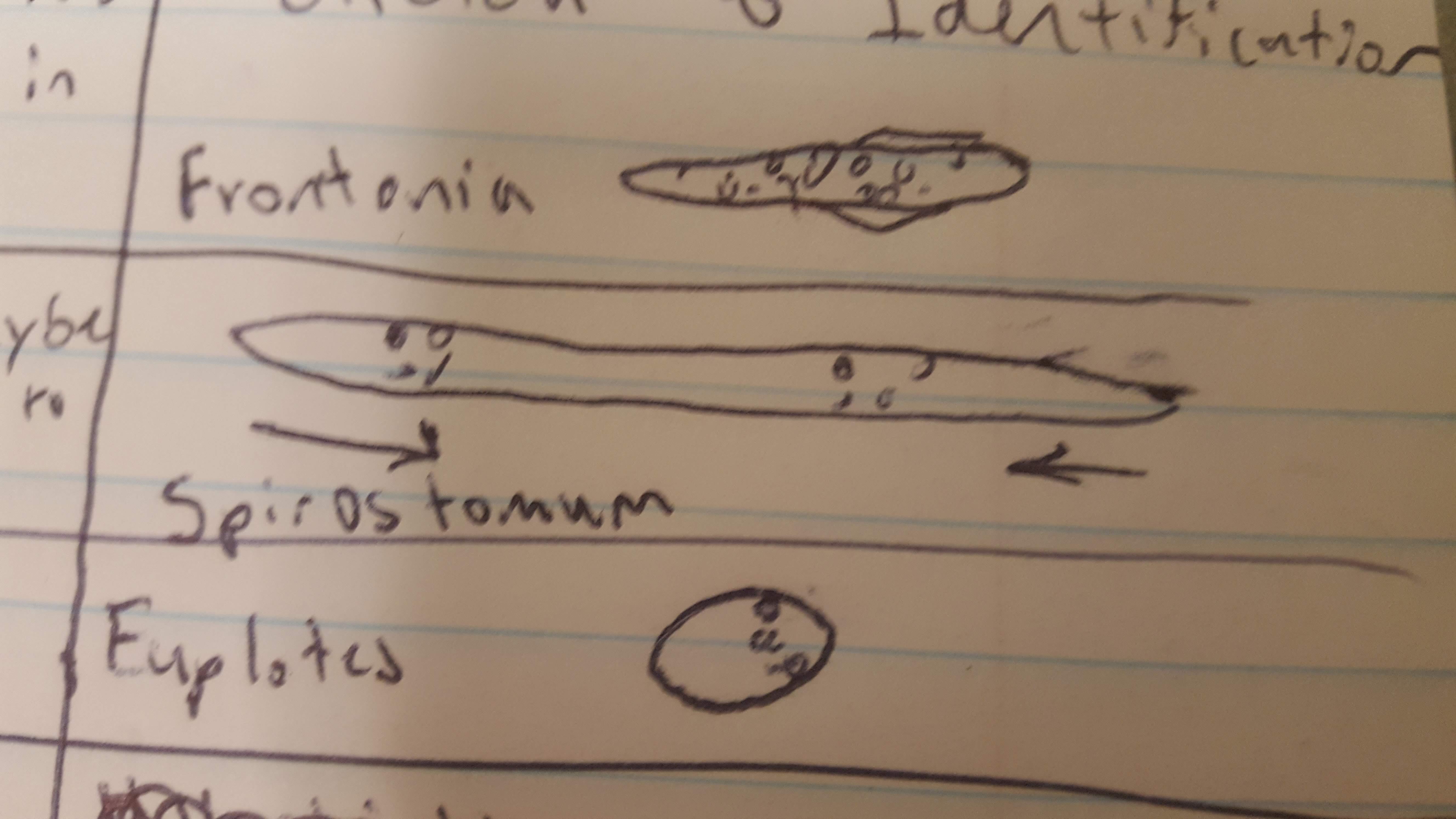
round with a crescent shape |
relatively large |
gilding while spinning |
short cilia, observable ‘organelle’ stretching from bottom around the edge of its body; located below surface |
Aspidisca |
| 3 |
‘cigar’ shaped |
very small |
very fast, spiral movement |
clear body with a single black dot on the tip; located on the surface |
Paramecium |
| 4 |
oval shaped |
relatively small |
medium speed, circular pattern of movement |
compressed itself to move, its movement was similar to that of a worm; located on surface |
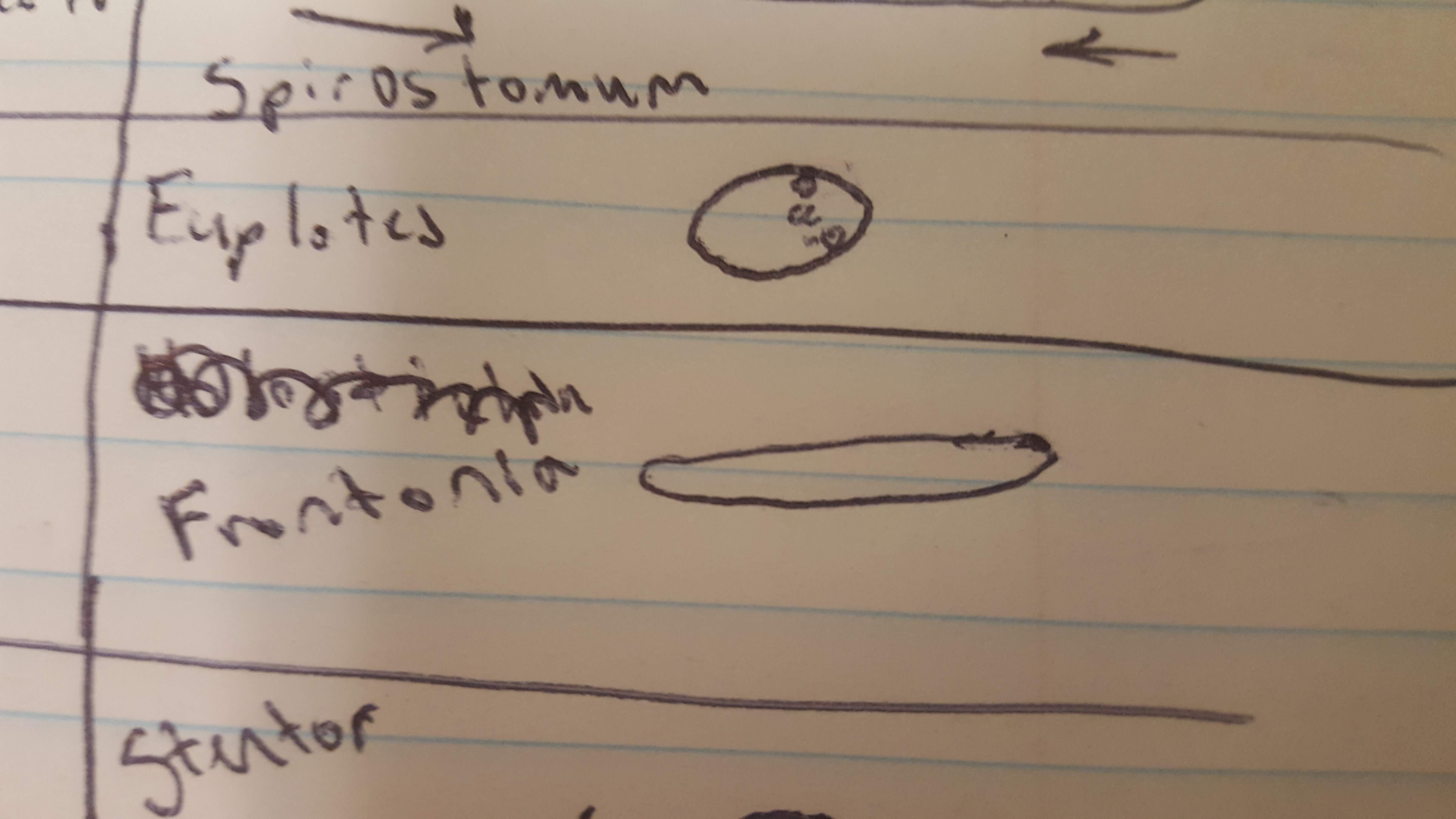
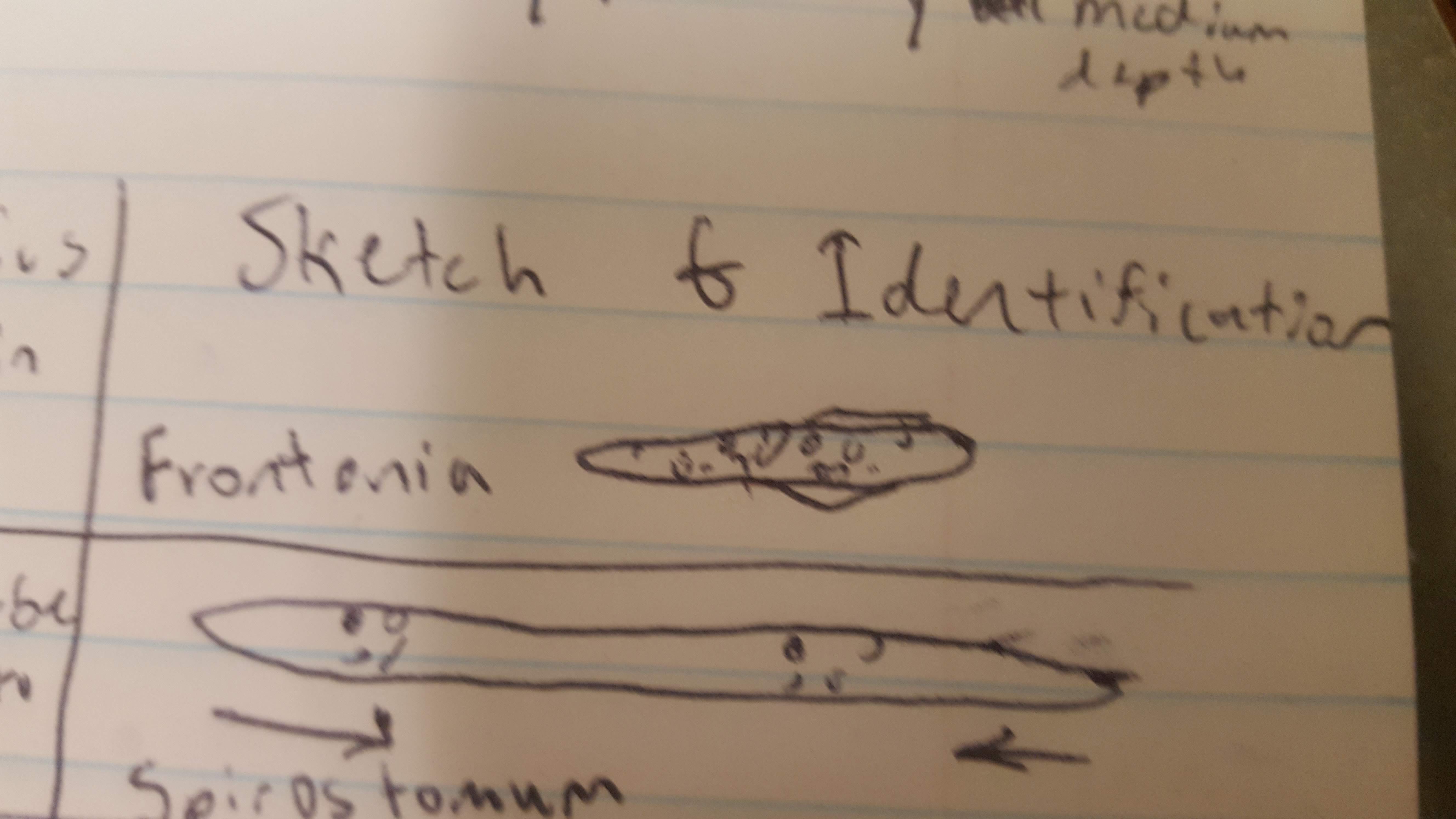
Frontonia |
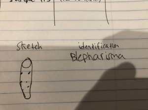
| 5 |
long narrow shape, bent at end |
very small |
contracted while moving, relatively slow |
brown color, transparent near front, black spots near end; located near surface; located close to surface |
Blepharisma |
| 6 |
cone, trumpet shape, large open front and narrow end |
similar size to #5 |
attached to material, fast when swimming freely |
very wide front; located on surface |
Stentor |
Storage:
Our samples were not stored and labeled.
Conclusions:
Using knowledge about ciliates, it is possible to identify them in a lab. Any error could have resulted from slight contamination between pipettes and test tubes, or from different lighting from the microscope. Other than that, there could be error from human observations, but since the identifications were mostly subjective anyway, this does not play as much of a role in this particular lab. I better understand the structures within cilates and how to use them to identify ciliates. I also am now more familiar with different types of ciliates than I was before conducting the lab. Moving forward, I know now that even if the results were not as expected or of the same format as expected, all results must be included and are still valid in the experiment. Despite being unable to identify unknown ciliate #1 due to its small size and fast speed, I was still able to include some results and my findings anyway. I now understand that labs are not meant to go exactly as expected, and that results are still valid even if different than expected. Now I will be able to be more open during labs, and more ready to accept whatever findings I have.


