April 25

Week 14

Print Friendly, PDF & Email

Objective

  • Compile all metadata into shared spreadsheet
  • Continue working on the scientific poster
  • Write an abstract

Procedure

  • Vote on a logo and shirt design
  • Finalize DNA samples
    • Collect sample ID and metadata on soil
      • Locations – coordinates
      • Tree species
      • Soil pH
      • Soil texture
      • ng/ul
      • Volume in tube
      • positive or negative PCR results
      • soil label
  • Work on poster
    • Address comments
  • Write final abstract
    • Use checklist provided on modules to write it

Abstract

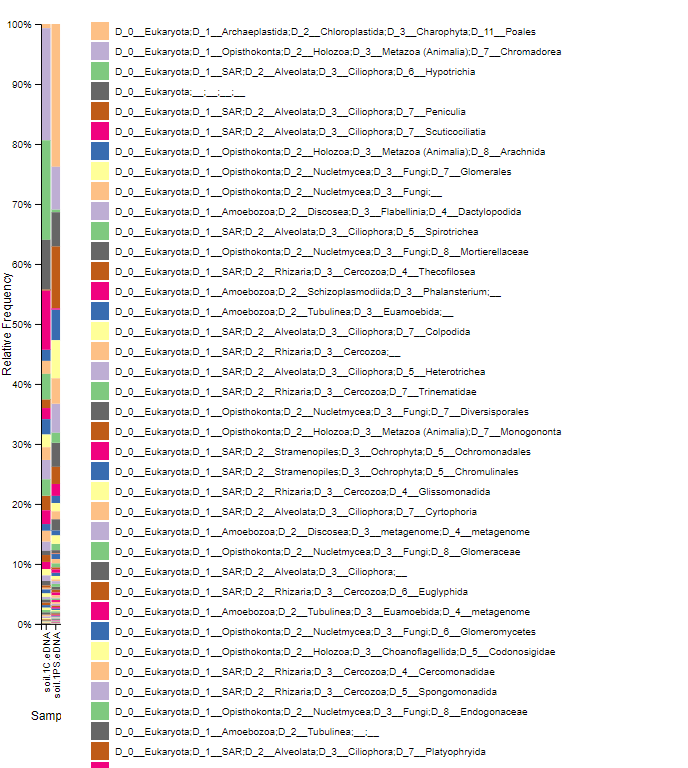
Soil ciliates have very little research done in its field, especially measuring the biodiversity of ciliates. Biodiversity of organisms, especially microorganisms in any ecosystem, is an important component to the stability of the ecosystem. Thus, this study was done in order to measure and quantify the measure of biodiversity of ciliates in soil ecosystems using a new method to extract eDNa, also giving us an idea if this new method, the silica bead method, is a viable one to extract eDNA in future studies. The silica bead method was utilized for the actual extraction of eDNA from the selected soil sample. Gel electrophoresis, the nanodrop spectrophotometer, and PCR were all effectively used to determine if the extraction of DNA from the soil sample was successful or not. After performing the eDNA extraction and the tests, the presence of DNA in our tests were negative, meaning we were unable to effectively extract eDNA from our soil sample. The DNA concentration was 7.1ng/ul as shown by the nanodrop spectrophotometer, an extremely low concentration. The gel electrophoresis fluorescent imaging found that there was no DNA in any of our lanes in both the pre-PCR imaging and the post-PCR imaging. The silica bead method was done correctly, without fail, leading us to analyze the metadata of our soil to determine if any of the other environmental factors prevented us from extracting eDNA. From the following results, we can reasonably conclude that there was no eDNA to extract from the soil sample or that there were certain factors surrounding the soil sample that prevented us from extracting the eDNA.

Poster

Conclusion

We were able to finish our abstract and scientific poster. Next week, we will be presenting our posters and demonstrating all the skills we learned over this semester.

April 18

Week 13

Print Friendly, PDF & Email

Objective

  • Continue using and getting comfortable with Qiime2
  • Analyze our eDNA from the soil samples, both the Chelex and PowerSoil samples, through Qiime tools

Procedure

  1. Retrieve the folder containing all the data from the Box link
  2. Open Terminal and make sure Qiime2 is installed and running correctly
  3. Activate Qiime2
    1. source activate qiime2-2019.1
  4. Change the directory to the downloaded folder in the Terminal
    1. cd (copy and paste the folder into the Terminal)
  5. Import sequences as Qiime2 artifact
    1. qiime tools import \

      –type EMPPairedEndSequences \

      –input-path emp-paired-end-sequences \

      –output-path emp-paired-end-sequences.qza

  6. Demultiplex the sequence
    1. qiime demux emp-paired \

      –m-barcodes-file sample-metadata.tsv \

      –m-barcodes-column BarcodeSequence \

      –i-seqs emp-paired-end-sequences.qza \

      –o-per-sample-sequences demux.qza \

  7. Create a summary for the demultiplexed sequences
    1. qiime demux summarize \

      –i-data demux.qza \

      –o-visualization demux.qzv

  8. Denoise up to 220 bases
    1. qiime dada2 denoise-paired \

      –i-demultiplexed-seqs demux.qza \

      –p-trunc-len-f 220 \

      –p-trunc-len-r 220 \

      –o-table table.qza \

      –o-representative-sequences rep-seqs.qza \

      –o-denoising-stats denoising-stats.qza

  9. Create a feature table to summarize and list all the sequences
    1. qiime feature-table summarize \

      –i-table table.qza \

      –o-visualization table.qzv \

      –m-sample-metadata-file sample-metadata.tsv

  10. Tabulate the feature table
    1. qiime feature-table tabulate-seqs \

      –i-data rep-seqs.qza \

      –o-visualization rep-seqs.qzv

  11. Visualize the stats of the denoising outcome
    1. qiime metadata tabulate \

      –m-input-file denoising-stats.qza \

      –o-visualization denoising-stats.qzv

  12. Create a phylogenetic tree
    1. qiime phylogeny align-to-tree-mafft-fasttree \

      –i-sequences rep-seqs.qza \

      –o-alignment aligned-rep-seqs.qza \

      –o-masked-alignment masked-aligned-rep-seqs.qza \

      –o-tree unrooted-tree.qza \

      –o-rooted-tree rooted-tree.qza

  13. Run the data against a reference database
    1. qiime feature-classifier classify-sklearn –i-classifier silva-132-99-515-806-nb-classifier.qza –i-reads rep-seqs.qza –o-classification taxonomy.qza
  14. Tabulate a table with all the sequences and classification information
    1. qiime metadata tabulate \

      –m-input-file taxonomy.qza \

      –o-visualization taxonomy.qzv

  15. Tabulate a barplot with all the classification information
    1. qiime taxa barplot \

      –i-table table.qza \

      –i-taxonomy taxonomy.qza \

      –m-metadata-file sample-metadata.tsv \

      –o-visualization taxa-bar-plots.qzv

Results

Demultiplexed:

Denoised:

Taxonomy Table:

Taxonomy Barplot:

Conclusion

I feel much more confident in working with Qiime2 and the Terminal. Running the commands is much more easier and I am beginning to understand what the different commands do and how the data is being processed.

April 12

Lab 12

Print Friendly, PDF & Email

Objective

  • Review the content from last lab
  • Complete the Moving Pictures Tutorial

Procedure

Moving Picture Tutorial

  1. Install Miniconda and Qiime following the instructions on the website and from last lab. On Terminal, after activating Qiime, input these codes in order.
  2. Create a new directory and change to that directory:
    1. mkdir qiime2-moving-pictures-tutorial
      cd qiime2-moving-pictures-tutorial
  3. Download the sample metadata:
    1. wget \
      -O “sample-metadata.tsv” \
      “https://data.qiime2.org/2019.1/tutorials/moving-pictures/sample_metadata.tsv”
  4. Make another directory:
    1. mkdir emp-single-end-sequences
  5. Download the sequence reads that will be used:
    1. wget \
      -O “emp-single-end-sequences/barcodes.fastq.gz” \
      “https://data.qiime2.org/2019.1/tutorials/moving-pictures/emp-single-end-sequences/barcodes.fastq.gz”
    2. wget \
      -O “emp-single-end-sequences/sequences.fastq.gz” \
      “https://data.qiime2.org/2019.1/tutorials/moving-pictures/emp-single-end-sequences/sequences.fastq.gz”
  6. Import sequence data in a different format:
    1. qiime tools import \
      –type EMPSingleEndSequences \
      –input-path emp-single-end-sequences \
      –output-path emp-single-end-sequences.qza
  7. Demultiplex sequences:
    1. qiime demux emp-single \
      –i-seqs emp-single-end-sequences.qza \
      –m-barcodes-file sample-metadata.tsv \
      –m-barcodes-column BarcodeSequence \
      –o-per-sample-sequences demux.qza
  8. Truncate sequences to 120 bases:
    1. qiime dada2 denoise-single \
      –i-demultiplexed-seqs demux.qza \
      –p-trim-left 0 \
      –p-trunc-len 120 \
      –o-representative-sequences rep-seqs-dada2.qza \
      –o-table table-dada2.qza \
      –o-denoising-stats stats-dada2.qza
  9. Tabulate metadata:
    1. qiime metadata tabulate \
      –m-input-file stats-dada2.qza \
      –o-visualization stats-dada2.qzv
  10. Create visual summaries of the data (feature table and feature data summaries):
    1. qiime feature-table summarize \
      –i-table table.qza \
      –o-visualization table.qzv \
      –m-sample-metadata-file sample-metadata.tsv
      qiime feature-table tabulate-seqs \
      –i-data rep-seqs.qza \
      –o-visualization rep-seqs.qzv
  11. Taxonomic analysis:
    1. wget \
      -O “gg-13-8-99-515-806-nb-classifier.qza” \
      “https://data.qiime2.org/2019.1/common/gg-13-8-99-515-806-nb-classifier.qza”
    2. qiime feature-classifier classify-sklearn \
      –i-classifier gg-13-8-99-515-806-nb-classifier.qza \
      –i-reads rep-seqs.qza \
      –o-classification taxonomy.qza

      qiime metadata tabulate \
      –m-input-file taxonomy.qza \
      –o-visualization taxonomy.qzv

    3. qiime taxa barplot \
      –i-table table.qza \
      –i-taxonomy taxonomy.qza \
      –m-metadata-file sample-metadata.tsv \
      –o-visualization taxa-bar-plots.qzv

Conclusion

This lab helped us learn and practice using the moving picture tutorial. It gave us images, charts, and data to help us understand our data better and analyze the data in an efficient manner. We learned how to use Qiime more, seeing how to manipulate the data using it and altering files to import the data into a physical image. For future steps, we will be using Atmosphere and Qiime to separate the ciliate DNA sequences to specific groups of ciliates.

April 4

Week 11: Poster Presentations (SW) Cloud Computing and Jupyter Notebooks

Print Friendly, PDF & Email

Objective

  • Visit a poster and “judge” it
  • Quick overview of CyVerse, Atmosphere, and Jupyter Notebook
  • Overview of Terminal

Procedure

  1. Meet at the computer lab
  2. Ask for access to Atmosphere on Cyverse
  3. Go over Qiime2
  4. Learn how to use the Terminal
  5. Install Qiime2 using the procedure laid out on the installation website
  6. Answer and finish the QTM

Conclusion

This lab was successful in that we set up the logistical side for future labs. For example, we went ahead and requested access to Atmosphere for Cyverse. We also went through what Qiime2 is and how this program is helpful for our future endeavors.

March 28

Lab 10: Next Generation DNA Sequencing

Print Friendly, PDF & Email

Objective

  • Learn about next generation sequencing and explain process of generating “reads” using Illumina sequencing
  • Cloud Computing: Set up your CyVerse account
  • Distinguish between an “image” and an “instance”

Procedure

  1. Meet at the computer lab
  2. Listen to small lecture going over the process of purified DNA to the final steps of sequencing
  3. NGS Illumina Card Challenge

Data/Results

The card challenge in the correct order.


Conclusion

This week’s lab is a great gateway into understanding the process of sequencing DNA and identifying the species in the soil samples we collected. A lot of new terms were given in this lab and as shown in the picture above, the work-flow of the whole process was taught.

March 21

Lab 9: Poster Presentations

Print Friendly, PDF & Email

Objective

  • Present scientific posters and improve them
  • Receive scientific and constructive criticism from not only just the faculty, but also from our peers

Procedure

  1. Meet at the computer lab
  2. Each group gave presentations on their current posters
  3. Each group received criticism on their posters and presentations
  4. The remaining time was used to continue to edit and improve upon the posters

Data/Results

This is our current rough draft of our scientific poster.

Conclusion

The biggest concern with our scientific poster is just content. Unfortunately due to circumstances, my group was unable to put more time into the poster than we had hoped to. However, we do have a focus for our poster. Because our results did not come out to be positive (negative results on the presence of eDNA from our soil sample), we are focusing on why that may be. In order to do so, we will be focusing on looking at the metadata between all the different groups and trying to pinpoint the different factors of why some groups had positive results and some had negative results.


March 7

Lab 8: PCR Results; Scientific Poster Design

Print Friendly, PDF & Email

Objective

  • Review over old journals, pre-labs, and QTMs
  • Run another session of gel electrophoresis on the amplified DNA

Procedure

Gel electrophoresis:

  1. Set the gel, already molded and made by the instructors, into the gel box
  2. Make sure that the buffer fluid is already covering the gel
  3. Load the wells with the ladder fluid and DNA
    1. 5 µl ladder fluid
    2. 10 mL DNA solution
  4. Run the gel for ~30 minutes at 100V

Results

This is the resulting UV light analysis of our gel. As shown by the above image, our DNA did not amplify through PCR very well or was present at all. Lanes 2 and 3 were my groups.

Overview

Today’s lab was very short in terms of work. We set up the gels and ran them. As they were running, we went and took a midterm quiz to review over what we have done so far in the course. Afterwards, we began the designing portion of our scientific powerpoint posters.

Future Steps

  • Continue to look over and review what we have learned over the course of this semester so far
  • Design our poster
February 21

Lab 6: Gel Electrophoresis and DNA Analysis

Print Friendly, PDF & Email

Objective

  • Determine if there is any DNA in our soil sample
  • Run gel electrophoresis and run the DNA through

Procedure

Gel Electrophoresis:

  1. Make sure the gel is placed in the buffer chamber in the correct orientation and covered with 1X TAE buffer
  2. Mix 9 µl of the DNA sample with 1µl of 10X loading buffer. Mix this tube
  3. Load 10µl of the sample into the second lane
  4. Load 5 µl of DNA mass STD 1 in lane 1
  5. Load 5 µl of DNA mass STD 2 in lane 3
  6. Run the gel at 100V for about 20-30 minutes

Imaging/Nanodrop:

  1. The gel was brought to an imaging lab and was placed in the MBC C305 to capture an image. The image was cast through UV lighting and looking for Ethidium Bromide.
  2. A drop of DNA samlpe was placed on the sampler to measure the absorbance.

Data

This is the result of the DNA imaging. It was set to look for Ethidium Bromide

This is the result of the nanodrop analysis of our DNA sample

ng/µl = 7.1

A260/A280 = 1.21

A260/230 = 0.25

The nanodrop did not produce the results that we were looking for.


Future Steps

Because the results were not the ones we were looking for, we may have to extract DNA again (Michael told us this might be the case). The results we got were disappointing, but are opportunities to grow and learn from.

February 15

Lab 5: DNA Purification and Gel Electrophoresis

Print Friendly, PDF & Email

Objective

  • Practice DNA purification on the eDNA we extracted from our soil samples
  • Set up gel electrophoresis tests
  • Begin setting up our methods for our research

Procedure

Chelex Procedure:

  1. Incubate sample for 30 minutes at 56°C on a heat block
  2. Broil for 8 minutes at 100°C on the heat block
  3. Vortex for 1 minute
  4. Centrifuge at 16000 xg for 3 minutes
  5. Transfer the supernatant to a clean tube and discard the pellet
  6. Store the tube in a freezer box

Agarose Gel Procedure:

  1. Make one liter of 1xTAE solution by adding 4mL of 10x TAE stock solution to 36 mL of DI water and add 0.4 grams of agarose.
  2. Swirl gently in a flask
  3. Heat the solution in the microwave for 2 minutes
  4. Swirl gently again
  5. Let it cool for a couple of minutes
  6. Add 0.002 micro-liters of Ehthidium Bromide to the solution to make it a 0.5 ug/mL concentration in the solution. Swirl gently
  7. Set up the gel electrophoresis box with rubber bumpers and then add the comb to make the wedges in the gel
  8. Add the solution to the box
  9. Allow the solution to solidify ~ 30 minutes
  10. After the solution solidifies, add a small amount of 1xTAE buffer over the gel
  11. Place the box in a plastic bag and store in the fridge

Conclusion

We were able to make a gel mold for our gel electrophoresis test and continued upon the Chelex protocal to finish extracting our DNA. The procedure for both protocols went smoothly. We also were able to check the soil sample cultures. These cultures were the same ones that we cultivated a couple of weeks ago when we were still trying to isolate ciliates from the samples.

Future Steps

The agarose gel we created was put in a bag and stored in the fridge. The DNA samples were also put in the freezer box. Next week, we will be continuing and go off of what we did this week.

February 7

Lab 4: DNA extraction

Print Friendly, PDF & Email

Objective

  • Analyze the soil texture of our soil samples
  • Check our cultured ciliates
  • Extract DNA from a soil sample and prepare it for purification (next lab)

Procedure

Finding and isolating ciliates:

  1. Using a micropipette, place drops of water from the prepared 24-well plate onto a concavity slide
  2. Under a dissecting microscope, search for any ciliates
  3. Make sure to inform the TA/Professor if any are found

Determining soil texture:

This is the soil texture triangle diagram. It is used to determine soil type through the percentages of the soil composition.

Extracting and Preparing DNA from the Soil Sample:

  1. Using a mortar and pestle, grind 1 g of the selected soil sample and 1 g of small silicon balls into fine powder
    1. My group selected Andrea’s soil sample
  2. Add 1 mL of DNA extraction buffer, the solution to break open cells without destroying DNA
  3. Add 10 mg of activated charcoal to get rid of the impurities in the sample
  4. Mix the buffer, charcoal, and soil sample together
  5. Transfer the contents into an eppendorf tube
  6. Incubate the tube at 65°C for 10 minutes in a water bath
  7. Centrifuge the tube, with the balancing concept in mind, at 12000g for 5 minutes at 4°C
  8. Transfer the supernatant to the microfuge tube

Data

Leaf Characteristics and Pictures:

After analyzing and referencing the “Figure of Leaves” resource, I determined that the tree is a Chionanthus virginicus. The reasoning is because the leaf contains the following features:

  • Elliptical leaf shape
  • The edges of the leaf is smooth; entire
  • Where the edge of the leaf meets the stem is acute
  • The tip of the leaf is also considered acute in shape
  • It is also palmately veined

Soil Texture:

The total measurement was 2.5 cm

Sand – 1.2 cm – 48%

Silt – 0.7 cm – 28%

Clay – 0.6 cm – 24%

After referencing the soil texture diagram, I determined that my soil sample was a sandy clay loam.

Ciliate Observations:

I did not observe any ciliates in my culture/24-well plate.

Unfortunately, I did not have the time to check my non-flooded plate.


Conclusion

It was unfortunate that I was not able to find any ciliates in my culture or non-flooded plate. With the new information I received, my soil sample being a sandy clay loam type, there may be a connection between the lack of ciliates and the soil type. It may be that the ciliates are unable to sufficiently survive in the soil composition that I had gathered for my soil sample.

Future Steps

We extracted our DNA this week, so I look forward to purifying them next lab. Dr. Adair will be bringing in the DNA purification kit, something I have never worked with/done before in my academic career.