Lab 4: Meet Tetrahymena

09/13/18
Haiden Jordal
Objective: Students will observe tetrahymena under different magnifications and microscopes. With further observations, students will be able to learn more about the model organism and how it functions. Students will also learn how to use micropipettes and use their primary source evaluating skills to find articles relevant to the research of tetrahymena.
Procedure and Materials:
- Practice using the micropipettes with disposable pipette tips using water and varying measurements.
- Obtain a tetrehymena sample in the 24-well plate.
- Observe the Tetrahymena in the well plate under the dissecting microscope.
- While looking through the microscope, pick up 5 ul of cells using a P-10 micropipette.
- Place the 5 ul of cells on a concavity slide and observe them under the compound microscope.
- Approximate the diameter of a cell with the knowledge of the FOV measurements.
- Record observations in lab notebook.
- Utilize the computer lab to brainstorm and research articles relating to possible experiments to be conducted between tetrahymena and microplastics.
Observations:
| Trials | Number of Cells in 5 ul | Approximate diameter of the cell |
| 1 (40x) | Over 100 | 60 um |
| 2 (100x) | 50 | 36 um |
| 3 (400x) | 20 | 30.1 um |
| Dissecting Microscope |  |
| Compound Microscope 40x | 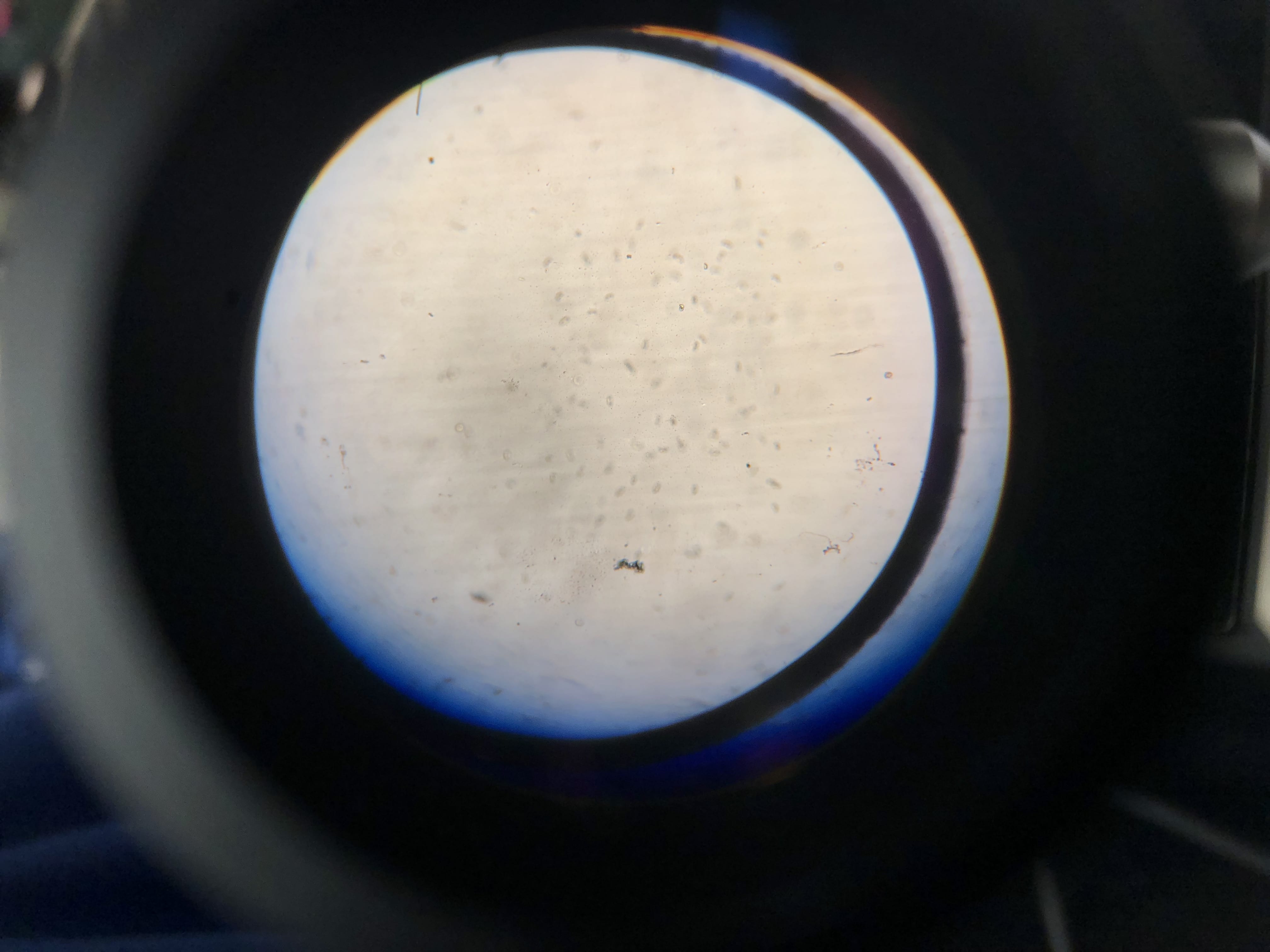 |
| Compound 100x |  |
Equipment Storage: Slides were cleaned and left to dry on the counter. Microscopes were covered after being set on lowest magnitude and stage brought up, and placed back in the middle of the lab table. The three ringed activity binder was placed back in the middle drawer. And all of the micropipette tips were disposed and the micropipettes were hung back on the rack in the center of the table.
Conclusion: Tetrahymena play an important role in biology and the study of genetics and many other cell functions. It is evident that the cells are easy to observe based on the amount of cells we could study under the microscope at one time. Also, since tetrahymena are easy to acquire, it makes replicating experiments and results quick and cost effective. With the different levels of magnification under the microscope and the FOV measurment calculated last week, I could approximate the diameter of one average tetrahymena cell to be around 30 micrometers(um).
Future Steps: With what we started in the computer lab, it will be important to study the tetrahymena more closely and accurately to be prepared to use them in our own experiments to understand the relationship between them and microplastics. Scientific literature will be a big resource to gaining the information necessary to understand their functions. In our experiment, we will be conducting trials of survival rates of tetrahymena that are exposed to soil contaminated with microplastics versus tetrahymena that are living in pure soil. The significance of this research is to find potential solutions to the looming threat of microplastic pollution to our biological ecosystem. With effective experiments we will be able to focus our area of study to wether or not microplastics will have a profound effect on soil organisms and wether or not that effect should be detrimental to the environment as a whole.