Lab 2 Post-Lab, The Ciliate Challenge 8/30/18

Objective:
The objective of this lab was to familiarize us to using the dissecting microscopes to observe microscopic organisms. The lab instructor had the goal for us to learn how to operate dissecting microscopes efficiently and being able to identify unknown ciliates by using information references and our basic knowledge of ciliates.
Purpose:
The purpose for this lab was to get us to understand the procedure of how to properly work in a lab and how set up our lab notebooks for recording data. Also this lab had the purpose to teach lab safety and how to use a dissecting microscope correctly.
Procedure;
- All students were placed in a team of three and each group were given 24 well plates, dissecting microscopes, six pipets and six unknown samples in closed test tubes.
- Each group member prepared their microscope by uncovering it and adjusting it to satisfy their preference in order for them to accurately observe the microscopic organisms.
- The group members then prepared their lab journal by making a chart of all the information they needed to collect for each of their samples. The information gathered for each sample included the organism’s number, shape, relative size, movement, extra characteristics, a sketch and identification.
- The group members divided the six samples between them, each member had to observe and identify only two samples.
- Each group member used aseptic techniques to prepare their sample for observing by carefully using a pipet to obtain a sample and place it onto one of the 24 well plates. All six of the samples were labeled with a number in order to prevent confusion.
- Each group member observed their sample individually and prepared their second sample with the same procedure used for first sample (step 5).
- While observing the sample, each group member noted all the information needed for identifying the ciliate.
- A reference guide of multiple ciliates was used to draw comparisons and help identify the samples.
- Lastly each group member sketched their sample and recorded their ciliate name once they identified it.
| Unknown # | Shape | Relative Size | Movement |
| 1 | Circular/round | Smaller than other cilialtes samples but narrow. | Very fast and quick |
| 2 | Round with a crescent shape | Relatively small and a similar to a circle. | Gliding while spinning |
| 3 | cigar shape | Relatively big | spinning/spiraling |
| 4 | Oval shape. Black dot near its front. | Relatively big with a large front compared to tail. | Large contractions when moving |
| 5 | long/narrow shape. End bends slightly | Small, less than 10% field of view. | Fast, worm like movement. Contrast slightly |
| 6 | Cone/trumpet shape. Large open front and narrow end. | Very small similar to size of #5 | Some were attached to plant surface. Others where fast and free swimming. |
#1
Sketch/Identification:
The subject was too small and fast to be able to accurately identified and sketched
#2
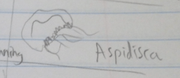
Characteristics- Short ridges on the bottom and has cirri to help with movement.
Identification: Aspidisca
#3

Characteristics: Large dot near the front that possibly could be an organelle.
Indentification: Paramecium
#4

Characteristics: small narrow shape with multiple small black circles that covered the inside of the organism.
Identification: Frontonia
#5
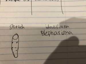
Characteristics: Brown color but had a white transparent circle on the front of the organism. Also within the organism were block circles that were identified to be organelles.
Identification: Blepharisma
#6

Characteristics:Unique trumpet shape with an opening on the front of the organism. The sample had a light blue and green mixture of color.
Identification: Stentor
Storage:
Our group cleaned the 24 plate wells by washing them with a bleach mixture and water. In order for the 24 plate wells to dry we placed them upside down above a piece of paper towel to avoid spilling water on the table. The six pipets and samples were then placed into the flume pit. The dissecting microscopes were put away by being covered and unplugged, the cord was wrapped around the arm of the microscope. Then we disinfected the table and dried it by using paper towels.
Conclusion:
In conclusion, this lab helped our group become comfortable with the microscopes and gave us a sense of how to properly work in a lab and use our lab notebooks. Our group was able to get a introduction into ciliates and began to understand some of their characteristics and their behavior. The first sample was not able to be identified but this was due to the subject being too small and fast that could not allow us to accurately identify it. Since this was our first lab and introduction to ciliates our identifications cannot be a hundred precent accurate but our group tried to the best of our ability to label these microscopic organisms. In the upcoming labs our group will have a better sense of a lab setting and will be able to work more efficiently than before.
Future steps:
For the future, our group could take less time trying to adjust the microscope to fit our needs since we gained beneficial information to handle the dissecting microscopes.