Lab 2: Ciliate Challenge 8/30/18

Objective of this lab:
The objective of this lab is to ensure that we are aware of how to use a dissecting microscope. By knowing this we can apply it to being able to clearly see ciliates through the scope.
Purpose of this lab:
The purpose of this lab is to view different ciliates through the dissecting scope and be able to identify what type they may be based on characteristics, shape, size movement and multiple other factors. In doing this we will gain knowledge on how to properly work the microscope and on the process of identifying ciliates.
Procedure:
- Uncover Dissecting Scopes and go over parts of microscope.
- Clean 24 well plate; Do this using a plastic pipette (each unknown goes in a separate plate no more than 1/2 full).
- There are 6 unknowns per groups of 3.
- Review 6 different unknowns and describe their shape, size, movement, location in media, characteristics and draw a sketch of the ciliate.
- Be sure to clean up your station. This includes cleaning the plates with bleach, cleaning the microscope and wiping the table.

Observations:
The six different unknowns all had unique characteristics that set them apart. This was helpful in identifying them. At first it was difficult learning how to focus the microscope in a way where you could clearly see the unknown. This was partly because the different unknowns floated at different levels in the liquid making it a constant point to re focus the microscope. Unknown 1 was oval like and relatively small. It moved somewhat quicker than the others and was located all over the media. It was clear with black points on the end and had a tadpole like shape. I identified it as possibly a paramecium. For this one the focus was at 2.6 Unknown 2 was circle like and small. It moved slowly in circles while also spinning. There was no specific location in the media, it was spread out. It was clear with a string like body and was also flat like. I identified it as possibly Aspidisca. For this one the focus was at 3.4. Unknown 3 was long and oval like and somewhat large compared to the others. It moved slowly and was spread out all over the media. It was long, dark colored and not as transparent as the others. I identified it as possibly Frontonia. For this one the focus was at 4. Unknown 4 was also oval like and long. It was small, moved in slow circles and was located all over the media. This unknown had a red tint to it. Identified it as possibly Spirostomum. For this unknown the focus was at 4. Unknown 5 was round and mostly in the center of the media. It was extremely small, black and moved slowly in circles. I identified this one as possibly being Euplotes. For this unknown the focus was at 3.5. The last unknown (6) was oval shaped and not flat. It was not small or large and did not move fast. It was free swimming and its presence was all over the media and it had a bluish tint. I identified this one as possibly being Stentor.
Storage:
We dispose of the ciliates in the sink and clean the small plates with 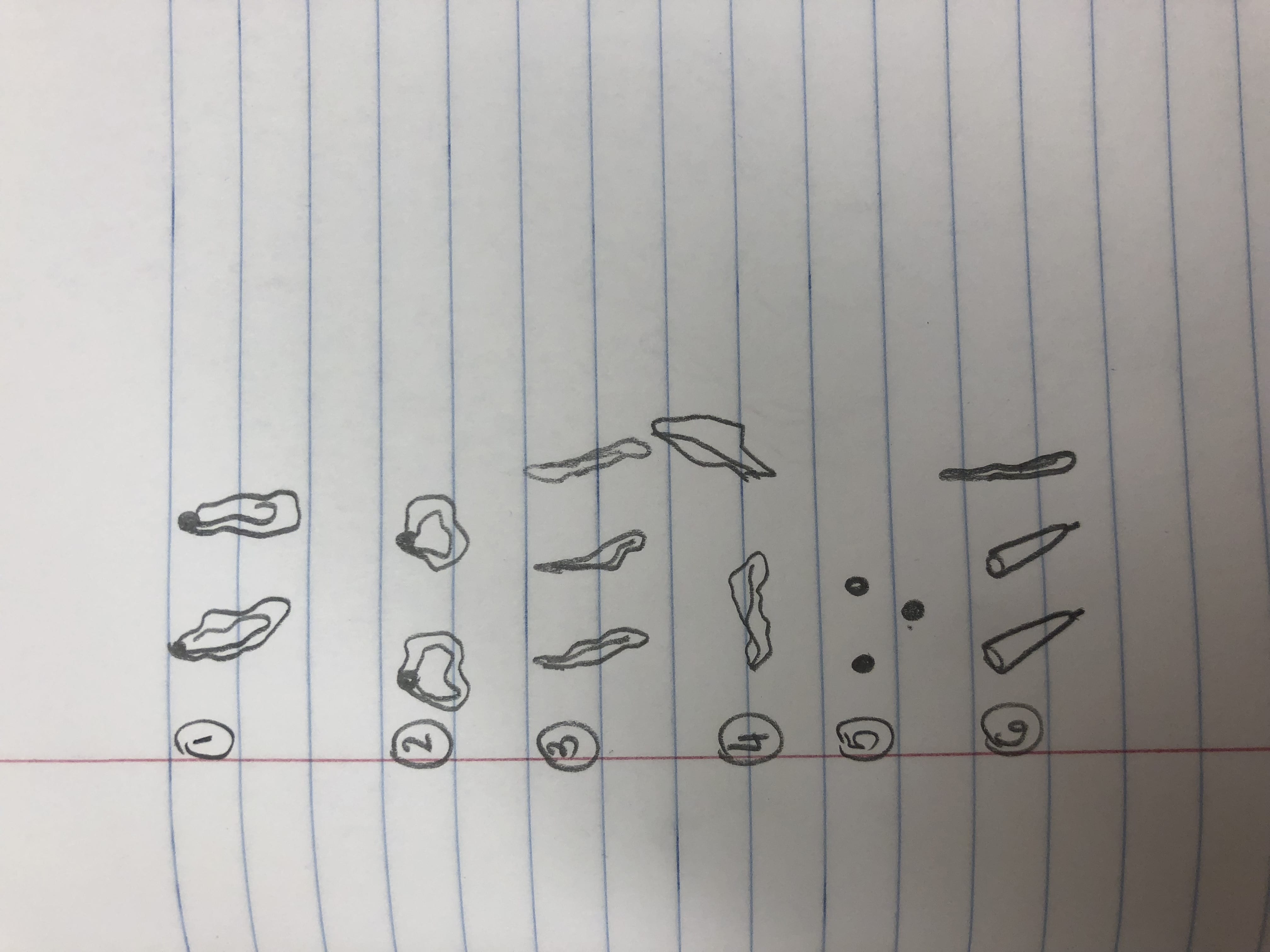 bleach. After this we left the plates on the left of the sink, right side down , to dry.
Future Goals:
In the future I want to be able to focus the microscope quicker. This will give me more time to be able to observe the unknown ciliates.