Lab #3: Primary Literature and Microscopy 9/6/2018

Rationale
In order to further understand ciliates, we learned to work with a compound microscope, concave slides, and stain. This exposed us to a ciliate’s reaction to stain and to the internal organelles that had been too clouded to see with the dissecting microscope. In addition to working the compound microscope and the parts associated with it, we also learned how to explore, discover, and scrutinize primary literary; specifically literature regarding the ciliate, Tetrahymena.
Procedure
Field of View
- Turn on compound microscope and focus on a prepared given slide on 4x, 10x, and then 40x magnifications. When focusing, make sure to use the coarse knob only on the lowest power and then proceed to use the fine adjustment knob until picture is clear.
- Once done familiarizing yourself with the compound microscope, place the slide to the side.
- Now, you will measure the field of view (FOV) diameter with a translucent ruler (to allow the light to shine through) on 4x magnification in millimeters (mm).
- Using that diameter and magnification, plug in the numbers into the following formula: FOVlow x Maglow = FOVhigh x Maghigh
- From this formula, calculate the FOV mm for 10x and 40x mag.
- Then, use the conversion factor 1000µm/ 1mm to convert the diameter in mm to diameter in µm.
- Record your values in a table.
Microscopy
- Now, grab a concave slide and slide cover.
- Procure a drop of a ciliate sample on the slide cover, making sure the drop is centered and only about a couple mm in diameter.
- Carefully, place the slide cover onto the concave part of the slide. It works best to approach it from an angle; this will result in a soft placement so as to not crush the sample.
- Once the slide is ready, begin observing it under the microscope.
- Record your findings in a table. I was not able to find any ciliates in my sample so the results in table 2 are my observations of my lab partner, Bella’s, ciliate sample.
- After observations are recorded and photos taken, allow your lab professor, TA, or LA to apply a stain to the ciliate.
- IMMEDIATELY observe the slide in order to see how the ciliate responds to the stain.
- Observe, record, and take pictures.
- Clean the slides and slide covers with water and place them on a paper towel to dry.
- Unplug, wrap up, and clean area.
Data
Table 1 FOV
| Objective Magnification | Diameter (mm) | Diameter (µm) |
| 4x | 4.5 | 4,500 |
| 10x | 1.8 | 1,800 |
| 40x | .45 | 450 |
Table 2 Pre-stain
| Description of shape and location of internal organelles | Short and skinny, organelles piled in the leading side, cilia not very visible |
| Color | Translucent |
| Movement | Constant and slow |

Table 3 Post-stain
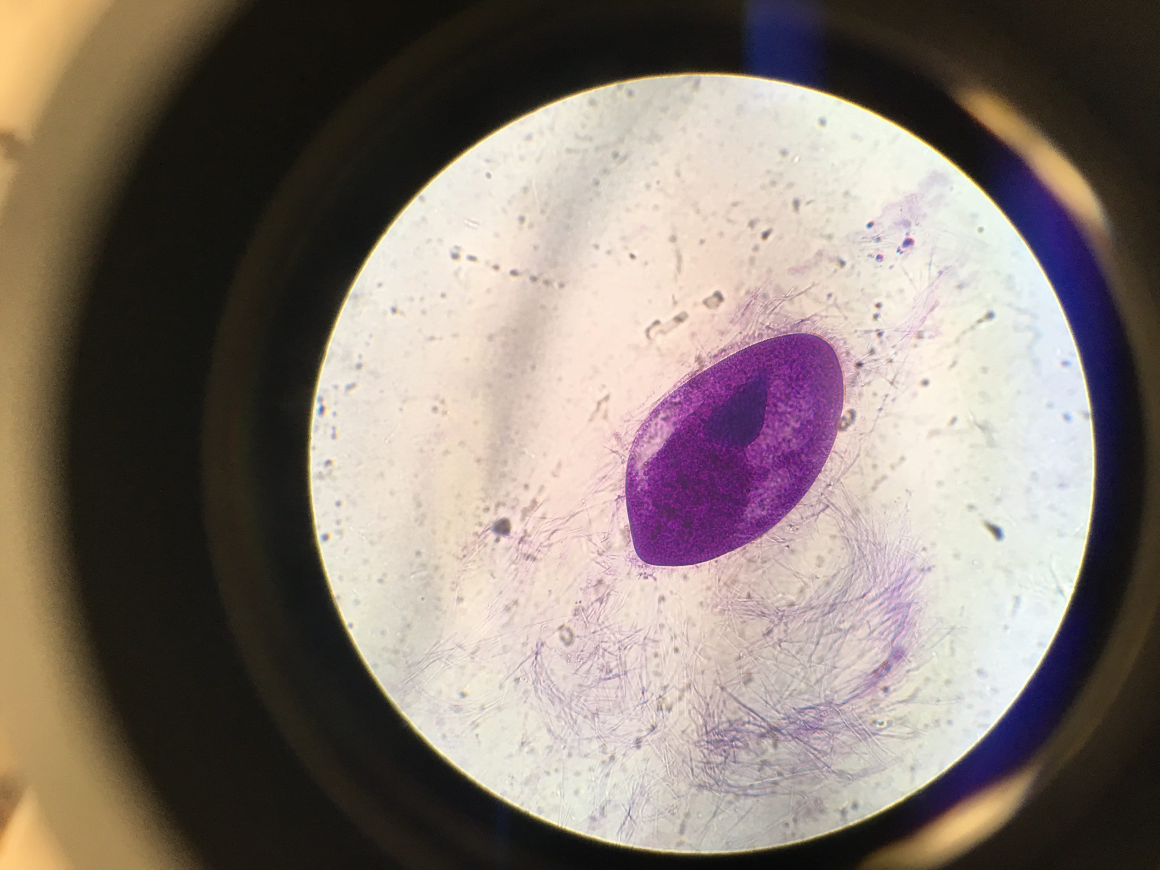
| Description of shape and location of internal organelles | Oval (resembling a football shape), all organelles packed in the center, cilia visible along the outside |
| Color | Purple |
| Movement | No movement |
Storage
As stated in procedure #9 under ‘Microscopy’, my slides and slide cover were cleaned off with water and placed on a paper towel to dry.
Conclusion and Next Steps
Today’s lab was exciting because we got to see more ciliates but this time, in different settings. The prepared slide given to us by our professor contained many ciliates that were easy to spot. Each ciliate had a different personality and it made for a great way to become comfortable with the compound microscope. However, when we were posed with the task to prepare our own slides, it became a bit more hands-on and by default, more difficult. I was able to expand my learning by having to take the information presented to me and apply it to my own slide preparation. Granted, the challenge proved to be a difficult one but this gave me an opportunity to work my lab partners by asking them if I could see their samples when my sample ended up being vacant of ciliates and how they successfully placed the slide cover onto the slide after I struggled getting the sample drop to remain in the concavity. This lab definitely showed me that science is not a single person practice, rather is takes a team to successfully explore different specimens.