February 26

02/22/18- Lab 7- CO1 PCR

Purpose

The purpose of today’s lab is to set up the gel for electrophoresis in order to prepare the DNA for amplification.

Procedure

Part 1: Introduction

Write down the general process for PCR.

Part 2: Prepare

  1. Obtain 3 tubes of pre-assorted Master Mix, which each contains 12.5 ml of 2x Master Mix.
  2. Label each of the positive control, negative control, and soil sample tubes.
  3.  Add 5 ul of the DNA template sample tube (10 nq/uL), and 5 ul of the DNA template to the Soil sample tube.
  4. Add 1 microliter of the cox1 primer to each of the tubes.
  5. Add 11.5 micro-liters of water to the negative control tube.
  6. Add 6.5 micro-liters of water to the soil sample test tube and the positive control test tube.

Part 3: Make 1.5% Agarose Gel

  1. Weigh out 0.6 g of agarose and add it to a Erlenmeyer flask.
  2. Add 40 ml 1xTAE to the Erlenmeyer flask.
  3. Cover the Erlenmeyer flask lightly with weighing paper and loose-fitting cap.
  4. Heat solution in the microwave until the solution is clear and small bubbles come off the bottom when gently swirled.
  5. Allow the solution to cool until the Erlenmeyer flask is comfortable to hold.
  6. Have your TA add 2 ul ethidium bromide and swirl gently.
  7. Assemble the electrophoresis box, making sure ends are sealed.
  8. Pour agarose smoothly into prepared mold, with as few bubbles as possible.

Data Collection

Part 1:


Above is the picture I drew explaining PCR. Nucleotides are always added to the 3′ end and two primers are used in the process.

Part 2:

Component Negative Control Volume Positive Control Volume Soil Sample Volume
2X Master Mix 12.5 ul 12.5 ul 12.5 ul
DNA Template 0 5 ul (10 nq/ul) 5 ul
Primers 1 ul 1ul 1 ul
Water 11.5 ul 6.5 ul 6.5 ul
Total Volume 25 ul 25 ul 25 ul

Our tubes were labeled +*, -*, and S*.

Part 3:

This was our solution before being heated. The solution is a cloudy white color and is a homogeneous mixture.

Both of the pictures above are the agarose in the electrophoresis apparatus. The solution is smooth with no air bubbles, which is great because it will prevent more issues when electrophoresis is carried out.

Conclusion and Storage:

The electrophoresis apparatus was left to be stored by Dr. Adair in the freezer. It is labeled LAK*. Overall, the lab was successful as we were able to prepare our sample for PCR. The hardest part was making sure that no air bubbles got in the agarose when transferring it to the electrophoresis apparatus. The next step is to proceed with amplification of the DNA through electrophoresis.


February 23

Lab 7: PCR Amplification and Ludox Extracted DNA

Purpose: To learn how to quantify DNA using the “nanodrop”, how to set up and run a PCR reaction, and how to make an agarose gel

Materials:

  • agarose
  • gel electrophoresis tub
  • TAE
  • microwave
  • cool bath
  • bromophenol blue
  • flask
  • bleach
  • Nanodrop

PCR workspace set up

  • Clean your desk with 10% bleach.
  • Wear gloves.
  • Clean/wipe down your pipettes before use.
  • Use autoclaved tubes and filter tips.
  • Keep all pipette tip boxes closed when not in use.
  • Keep all tubes closed when not in use.
  • Minimize the movement/ air flow around your desk.

PCR Reaction Set Up

Perform PCR assay in a total of 25 µL:

1. Add 12.5 µL  2X Master Mix to a flask. Add 0.6 grams of agarose to the solution and dissolve the substance at 45C for annealing.

2. Add 0.6 grams of agarose to the solution and dissolve the substance at 45C for annealing. (the mixture needs to be 1.5% agarose)

3. Allow the solution to cool before inserting 5ul of bromphenol blue in the agarose solution

4. Pour the agarose solution into the electrophoresis tub and allow it to sit still in order to solidify. The next step will be to insert the DNA into the gel.

Conclusion

The lab went smoothly however, I am still a little worried about the amount of DNA that will be present in our sample due to the mishap in the previous lab. After the DNA is placed in the gel, it will move towards the positive side of the tub since DNA is negatively charged. The strands will have to be observed under a UV light, this was the point in adding the bromothenol blue so the DNA will be clearly visible.


February 23

Lab 7: PCR Amplification and Ludox extracted DNA 02/22/18

Purpose/Objective

The purpose of today’s lab was to prepare DNA for amplification and to begin setting up our agarose gel for gel electrophoresis. The lab introduced us to putting together all of the primers, water, DNA, and buffers into a single solution to prepare to DNA Amplification.

Procedure

Preparing DNA Tubes

  1. Obtain 3 tubes which are pre-filled with 12.5 μL of 2X Master Mix. Label the tubes so that you know which tubes will serve as your positive and negative controls, and which will serve  as your sampled DNA tube.
  2. Add 1 μL of COX1  primers,  and 11.5 μL of water to the negative control tube.
  3. Add 1 μL of COX1 primers, 5 μL of Paramecium DNA, and 6.5 μL of water to the positive control tube.
  4. Add 1 μL of COX1 primers, 5 μL of sample DNA, and 6.5 μL of water to the sample DNA tube.
  5. Store the tubes away for next week.

Preparing Agarose Gel

  1. Measure out 0.6 grams of Agarose powder on a scale. Empty the powder into an Erlenmeyer flask.
  2. Measure out 40 mL of T.A.E. and empty into the Erlenmeyer flask. Swirl the flask to thoroughly mix the solution.
  3. Cover the top of the Erlenmeyer flask with weighing paper and a loose-fitting cap, and microwave the solution for 1 minute and 20 seconds at 70% power.
  4. Remove carefully, and allow to cool in a water bath at 55 degrees Celsius for 5 to 6 minutes.
  5. Add 2 μL of ethidium bromide to the solution. This will cause the solution to bind to the DNA.
  6. Pour the solution into a gel rack on a flat surface to cool and solidify into gel.
  7. Clean lab area and say bye to Will.

Conclusion

This lab was a pretty productive lab that was also fun. We didnt’ have any observable data for this lab, as all we did was prepare our DNA tubes and agar gel for next lab. Next week I believe that we will pour our DNA onto the gel, and run a 100 Watt  electrical current through the gel. Apparently the DNA is supposed to show up using gel electrophoresis. I think the only error that may be made was pipetting such small amounts of the primers into the tubes. I feel like it was hard to tell if your pipette was actually extracting and releasing the 1 μL of COX1 primers into the tubes but we will see next class.

February 23

Lab 7: Cytochrome Oxidase 1 (cox1/COI) and PCR 2/22/18

Objective:

In lab our goal is to learn how to quantify DNA using the “nanodrop” technique, how to set up and run a PCR reaction as well as learn to make an agarose gel. We hope to successfully complete the PCR reaction after learning the procedure, and achieve the end goal of being able to complete the metabarcoding process and be able to analyze as well as classify soil ciliates.

Purpose:

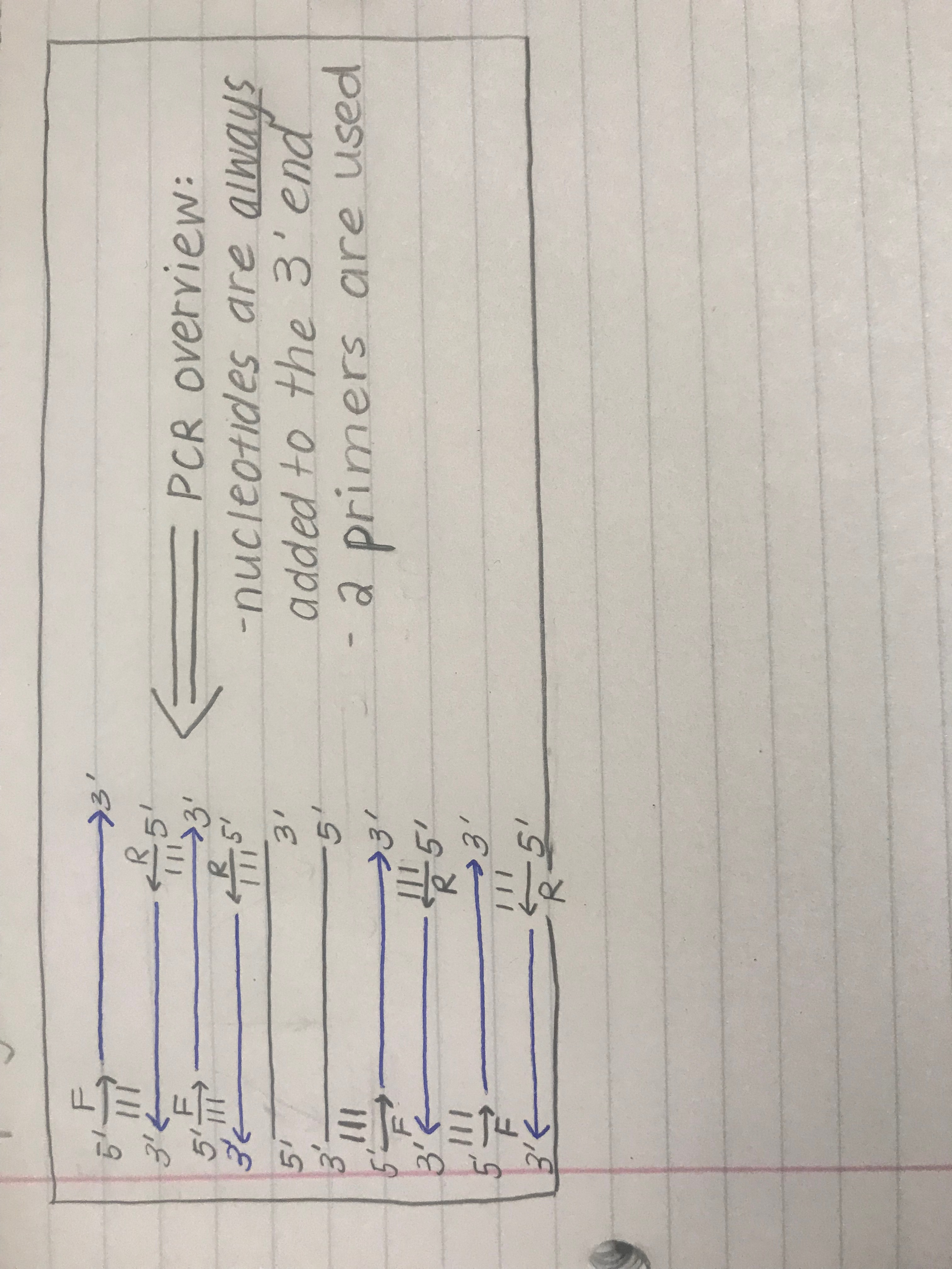
The purpose of today’s lab is to continue the PCR process and testing positive and negative controls as well as the DNA extracted from the soil sample. The purpose of learning how to make a gel and actually physically creating it, is to be able to successfully run gel electrophoresis. After reading the scenific literature provided from our pre-lab, we are able to conceptualize how the cox1 gene primers bind to the DNA and how this will allow us continue in our metabarcoding process.

Procedure:

*Use bleach to create a sterile work station. This will help avoid any contamination from other microbes and bacteria on the lab tables.

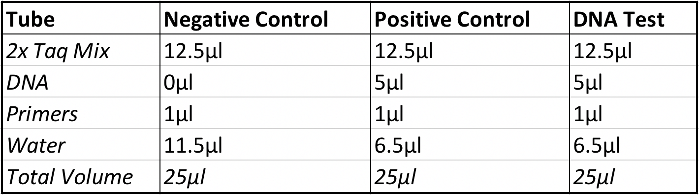
1.) Obtain three small tubes containing 12.5 micro-liters of the TAQ polymerase mixture.

2.) Label the tubes, “DNA test”, “positive control”,and “negative control”.

3.) Add 5 micro-liters of the DNA template sample to the tubes labeled “DNA test”, and “positive control”.

4.) Add 1 microliter of the cox1 primer to each of the three small test tubes.

5.) Add 11.5 micro-liters of H2O to the negative control tube.

6.) Add 6.5 micro-liters of water to the DNA test tube and the test tube labeled positive control.

7.) Add up the volume of everything added into the tubes to ensure that each has a final volume of 25 micro-liters.

8.) Place the tubes in a tube rack at the front of the room, record the place of each of the rubes within the rack.

9.) Store the tubes at 4 degrees Celsius or freeze until confirmation through gel electrophoresis may be performed.

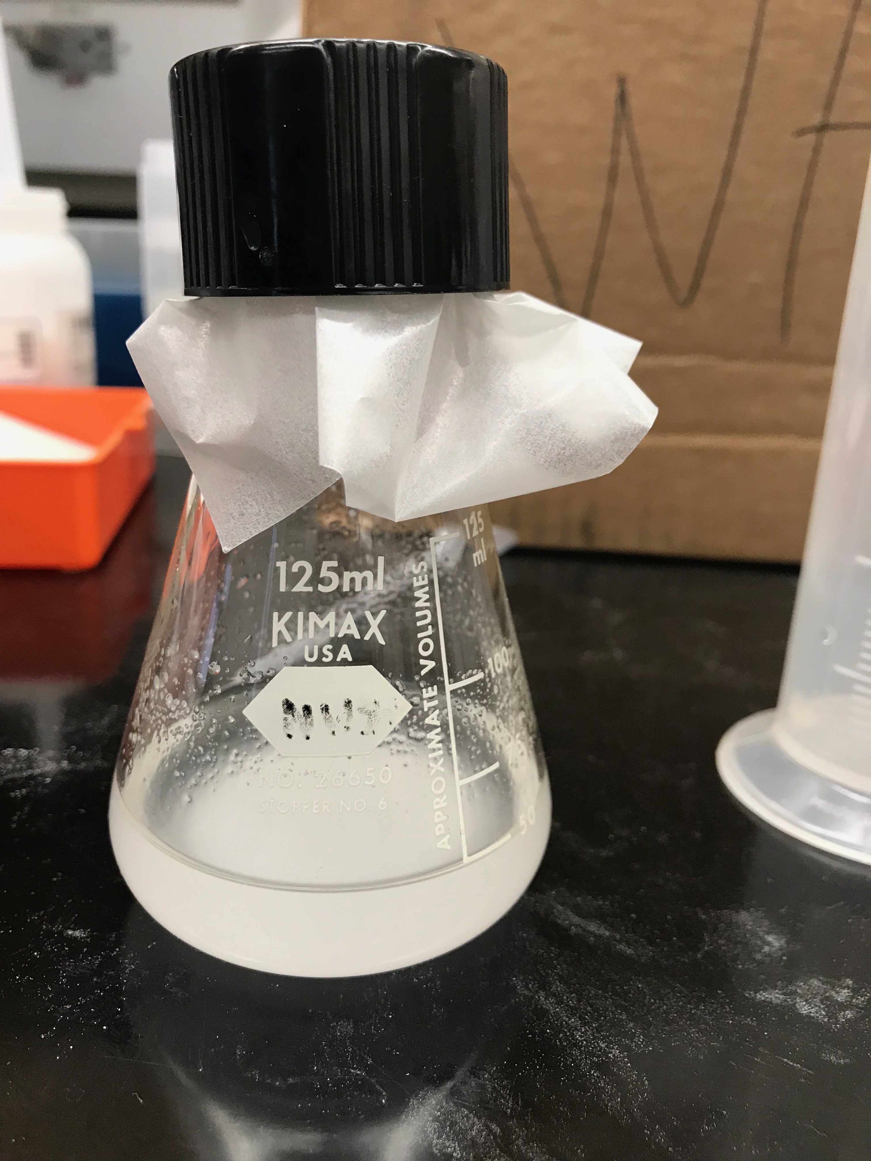
10.) Proceed to making the gel for gel electrophoresis by adding 0.6 grams of agarose to 40mL of 1 x TAQ in an erlenmeyer flask. (Weigh the agar on a scale using weigh paper, tare the balance after placing the weigh paper on the scale and add agar powder until there is 0.6 grams).

11.) Cover the top of the flask with weigh paper and then place the lid loosely over both to prevent it from exploding in the microwave.

12.) Place in the microwave at power 7 for 1 minute and 20 seconds, or until boiling.

13.) After, place in heat bath for about 5 minutes then add ethidium bromide.

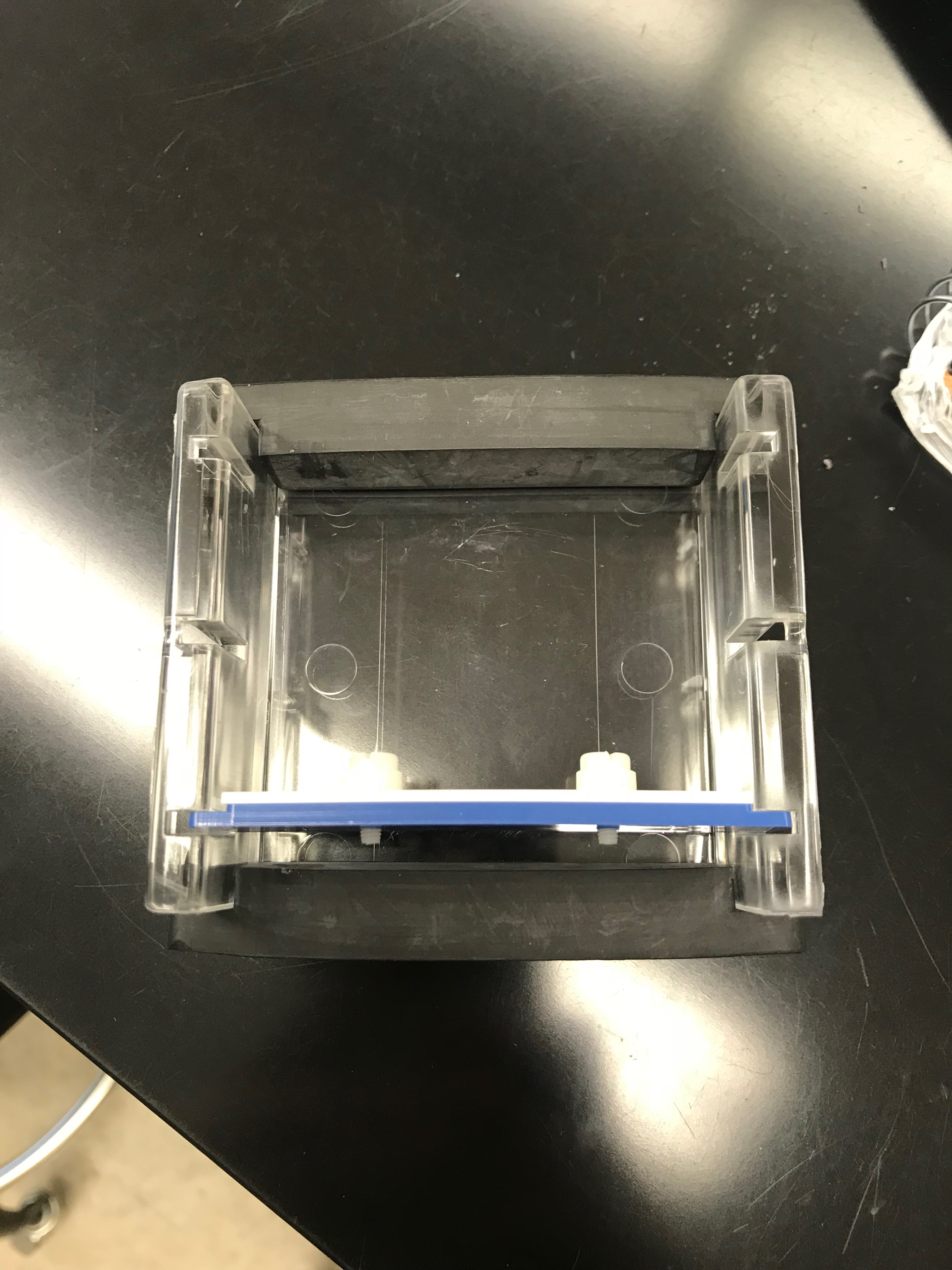
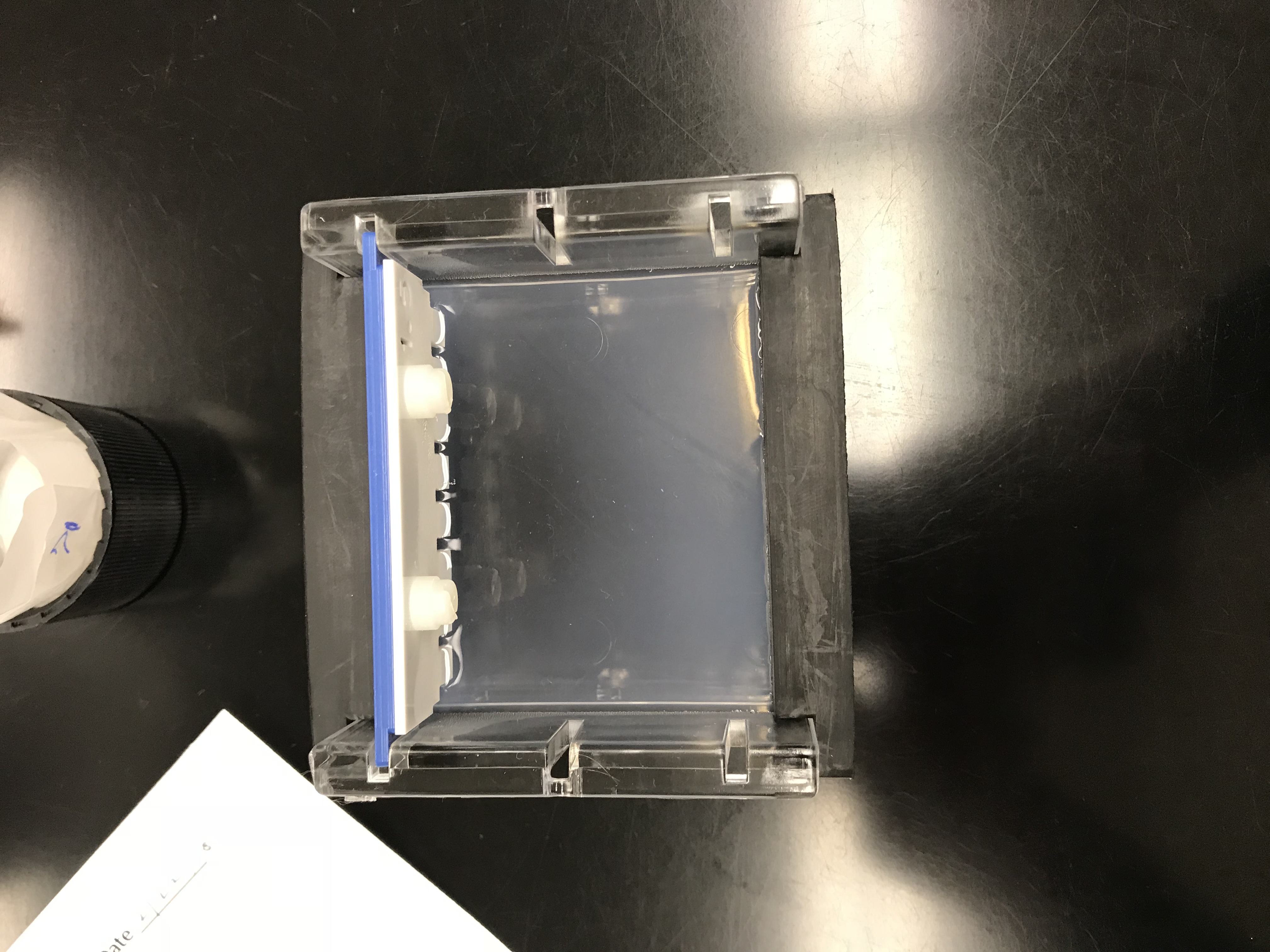
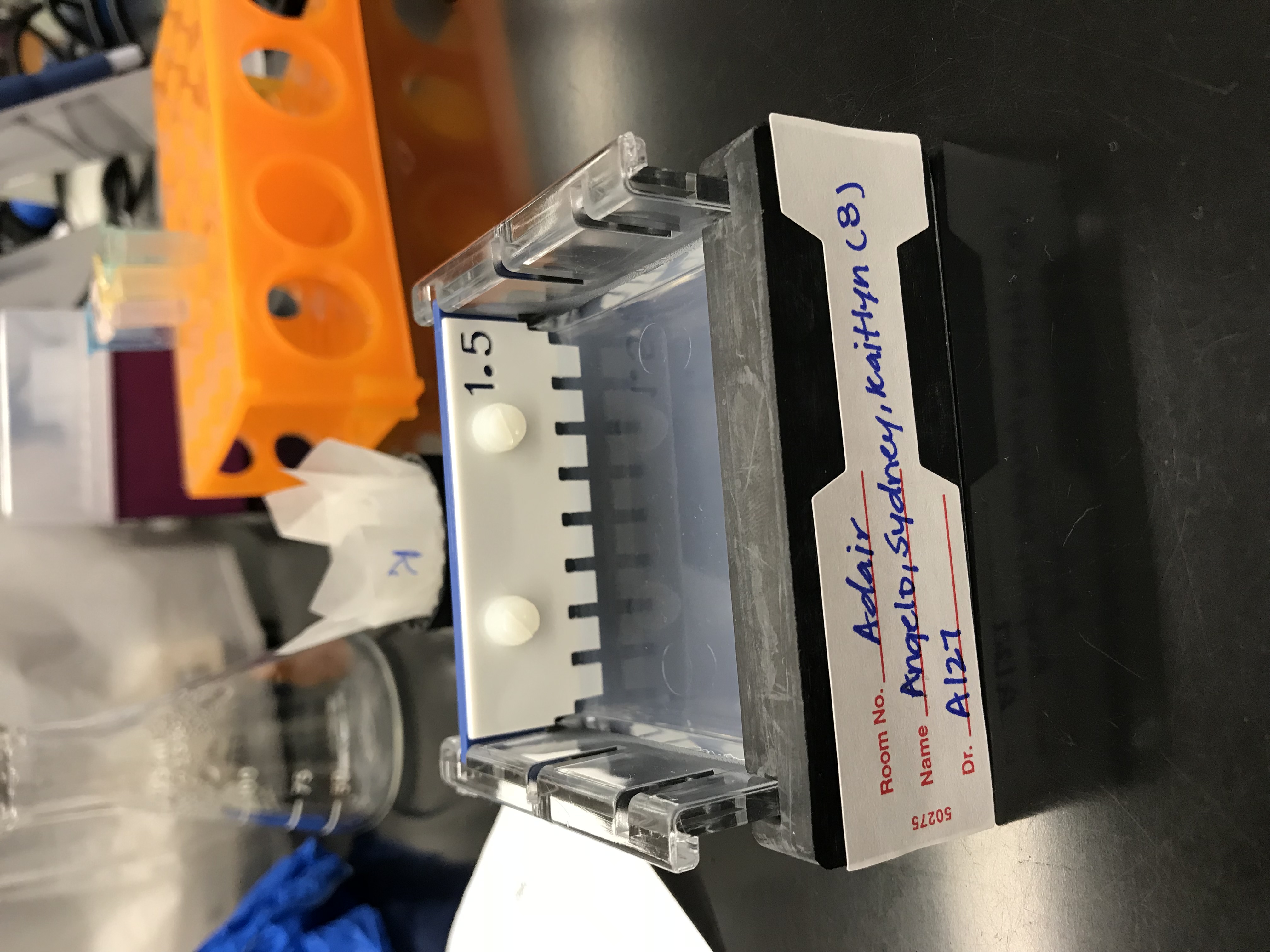
14.) Pour the mixture in a gel mold and label is with your group identifier (LSK G#5).

15.) Let sit for 30 minutes then store in the refrigerator for next lab.

Data/ Observations:

COX 1 Gene Forward Primer:
5′-ATGTGAGTTGATTTTATAGAGCAGA-3′

COX 1Gene Reverse Primer:
5′-GGDATACCRTTCATTTT-3′

PCR Cycling Conditions for COX 1 Primers:
Stage 1: 94 degrees Celsius for 4 minutes.

Stage 2: 5 cycles of denaturation at 94 degrees Celsius for 30 seconds, annealing at 45 degrees Celsius for 1 minute and extension at 72 degrees Celsius for 105 seconds.

Stage 3: 35 cycles of denaturation at 94 degrees Celsius, annealing at 55 degrees Celsius for 1 minute and extension at 72 degrees Celsius for 105 seconds.

Stage 4: Hold at 72 degrees Celsius for 10 minutes.

*We were required to where gloves for sterilization purposes and because ethidium bromide is a carcinogen.

*After pouring the gel into the mold, we believe that the liquid did not distribute equally before it started to harder, causing it to have a sense dense area of the agarose mixture around the wells.

Storage:

The three small tubes with the DNA test, positive and negative controls, were placed under the projector in a tube rack labeled on one axis with numbers and the other axis with letters. The DNA test tube is located in slot B1 labeled “DNA” on the side and a “5” that represent group number five. The positive control tube is located in slot B2 and is labeled with a “+” on the side and a “5” on the top and the negative control tube is located in slot B3 and is labeled with a “-” on the side and a “5” on the top. After creating a gel, we let it sot for thirty minutes so it could take the shape of the mold. It was later left on the end of our lab tables to be picked up and stored in the refrigerator so that we may use it during the next lab. The gel mold has a piece of blue tape that is labeled “LSK G#5” which stands for “Lindsey, Sandi, Kaitlyn Group # 5” which we have created as our group identifier.

Future Goals:

After completing the steps of PCR and creating a gel, I hope that we are able to run gel electrophoresis and are able to view the DNA fragments under the UV light. Our gel was not evenly distributed throughout the mold; I hope that this will not effect the DNA fragment traveling through it. I am somewhat worried that we will not be able to clearly see and DNA markers on the ladder in the gel because of our soil samples. In the future, I would like to be able to continue on with the metabarcoding process and hope the see the DNA fragments with in the gel so we can compare it to the markers of other organisms and classify them.

February 23

Ludox Protocol Completion & EZNA DNA Protocol Lab

February 22, 2018

Purpose:

The purpose of this lab was to learn about the process of PCR and carry out the beginning of the protocol with our soil DNA samples and a positive and negative control. Students are also to make agarose gel and construct the gel electrophoresis apparatus for later use.

Procedures:

Part 1:

  1. Discuss learning objectives with class.
  2. Draw out process diagram for PCR.

Part 2:

  1. Prepare lab for PCR by wiping down the lab tables with bleach, tying back hair, and putting on gloves.
  2. Obtain 3 tubes of pre-assorted Master Mix (12.5 uL in each)
  3. Label each tube on the side with a sharpie (+,-, S)
  4. To the negative tube, using a p10 micropipette add 1 uL of the primers (COX 1 forward and reverse), and 11.5 uL of water (6.5 uL first followed by 5 uL for a total 11.5 uL).
  5. To the positive tube, using a p10 micropipette add 1 uL of the primers(COX 1 forward and reverse), 6.50 uL of distilled water, and 5 uL of the control DNA.
  6. To the tube labeled S, using a p10 micropipette add 1 uL of the primers(COX 1 forward and reverse), 6.50uL of distilled water, and 5 uL of the soil DNA.
  7. Label your group number on the top of each tube with a sharpie.
  8. Place the 3 tubes in the tube rack at the front of the classroom making note of the location you put it in. Record number/letter placement label on QTM sheet.

Part 3:

  1. Using analytical balance, weigh out 0.6 grams of agarose, and add it to Erlenmeyer Flask.
  2. Add 40mL of TAE (accurately measured with a graduated cylinder)  to the flask.
  3. Close the flask with paper and stopper.
  4. Microwave it for 1 minute and 20 seconds at a power of 7 making sure it is boiling.
  5. Place the flask in a 55 degrees Celsius water bath for 5 minutes.
  6. Add 2uL of ethidium bromide and mix.
  7. Assemble electrophoresis apparatus, making sure there are no openings and pieces are compacted.
  8. Slowly add the gel to the apparatus, and allow it to cool for 20 minutes before storing in the freezer.


Data:


Negative Control Volume (uL) Positive Control Volume (uL) Soil DNA Volume (uL)
2X Master Mix 12.5 12.5 12.5
DNA 0.00 5.00 5.00
Primers 1.00 1.00 1.00
Water 11.5 6.50 6.50
Total 25.0 25.0 25.0


The initial concentration of the stock primer was 10 uM.

The final concentration in our sample was 0.4 uM.

Tubes placed in A1, A2, A3.


Conclusion:

This lab was successful as we were able to prepare our trial samples for PCR. Our results from last week did not come out the way we had hoped (NanoDrop curve was significantly off) so we were unable to get an estimate at the concentration of DNA we had in our sample. Most of the components were already pre-assorted by the lab team, so it was easy because we were just adding things to the mix. The most difficult part of the lab was making sure that we were keeping a sterile environment. We did this by wearing gloves, cleaning out tables with bleach, making sure we weren’t speaking in the direction of the samples, and making sure the pipette boxes and tubes were being closed. The next step is to run our trial samples under current in gel electrophoresis to make sure we get bands. We will then send our samples to a lab for analysis and sequencing. In the future, it will be beneficial to have half the class doing the protocol in a slightly different way so we have a better chance of getting positive results.

Gel Apparatus labeled: Group 7 & Stored: in freezer

February 23

Lab #7-PCR DNA Amplification and Gel Electrophoresis (2/22/18)

Purpose:

The purpose of this lab was to prepare positive and negative control samples and an eDNA soil sample for PCR amplification by adding 2X Master Mix, DNA template (only to the positive control and soil sample), water, and the primer COX1. The PCR Amplification process includes denaturation, annealing, and elongation. In addition, we also prepared the gel needed for gel electrophoresis.


Procedures:

  1. Sterilize the work environment by cleaning the table with bleach.
  2. Add 2X Master Mix, DNA template, water, and primer to the designated tubes so that the volume of each tube is 25uL. (Note: See the table in “data/observations” for specific quantities.)
  3. Add 40mL 1xTAE to 0.6g agarose in a small Erlenmeyer flask.
  4. Cover lightly with weighing paper/Kimwipe and loose-fitting cap (Note: Do NOT tighten tightly close the cap otherwise an explosion might occur.)
  5. Heat until solution is clear and small bubbles come off the bottom when gently swirled.
  6. Allow to cool until the flask is comfortable to hold (5-6 minutes).
  7. Have your TA add 2uL ethidium bromide, swirl gently.
  8. Set up gel electrophoresis box, making sure the open ends are sealed.
  9. Pour agarose gel smoothly into prepared mold, with as few bubbles as possible. Insert the comb with its back towards the nearest edge. Allow it to solidify for at least 25-30 minutes.
  10. Cover gel with prepared 1xTAE buffer solution so that it will not dry out.
  11. Carefully remove comb and turn gel so that the wells are furthest away from positive electrode (think “run to red–you want the negative DNA to run towards the red positive electrode.)
  12. Use a micropipetter, add 5uL of the ladder and 10uL of each pCR product + loading buffer. If the loading buffer is not included in the Taq polymerase used in the PCR, add 5uL loading buffer to the PCR product and mix thoroughly before transferring to the gel.
  13. After you have loaded your sample, place the lid on your box and turn on the power supply to approximately 100 volts. Allow to run for 30 minutes or more, allowing the loading dye to run approximately halfway across the gel before turning off the power.
  14. Image with UV light.


Data/Observations:

Calculation determining the amount of agarose:

X g agarose / 40mL buffer = 0.015

X = (40mL)(0.015)

= 0.6 g agarose

Each tube (from step 2 of the procedures) is labeled with a “6” on the top and a “+”, “-“,  or “s” on the side. Our gel rack is labeled “6 LBP”

Component

Volume (Neg. Control Tube)

Volume (Pos. Control Tube)

Volume (Soil DNA Tube)

2X Master Mix

12.5 uL

12.5uL

12.5uL

DNA Template

0

5uL

5uL

Primers

1.0uL

1.0uL

1.0uL

Water

11.5uL

6.5uL

6.5uL

Total

25.0uL

25.0uL

25.0uL


Conclusion:

This lab was great exposure to the PCR amplification process and was the next step in allowing us to classify ciliates based on their genetics. I thought it was really cool that we we were able to make our own gel and that this protocol was fairly hands on. I am excited to run the gel electrophoresis next week and to finally be able to analyze the results of our organisms.

February 23

Lab 7: PCR and Gel Electrophoresis (02/23/18)

Purpose:

  • The objective of the lab was to prepare the DNA for amplification. by making a positive and negative control along with a soil sample one. Also we started the process of gel electrophoresis by creating the gel agarose and storing it for the next lab once we continue.

Procedure:

  1. Before we began the process, we made sure to wash our work area with 10 % bleach
  2. The researchers were given a sheet that had a table of values of the proportions of water, primers, 2X master mix, and DNA we need to pipette into a mini tube.
  3. These values were different for each of the groups that we had: positive control, negative control, and our soil sample.
  4. Once we included the respective proportions of contents in each of these tubes, we made sure to label the mini tubes with our group number and placed on the rack with the rest of the class.

Preparing the Agarose Gel

  1. First we determined how many grams of agarose powder we need for a 1.5% solution of agarose T.A.E. by multiplying 1.5 with 40 mL of T.A.E.
  2. We determined the mass of agarose needed was approximate 0.6 grams.
  3. The 0.6 grams of agarose powder was then transferred to an Erlenmeyer flask and 40 mL of T.A.E. was then put in the flask also and sealed by wax paper.
  4. The flask was then swirled and inserted into a microwave where it was in there for 1 minute and 20 seconds under 70 power.
  5. After it was taken out of the microwave, we carefully placed it in a water bath for 5 minutes.
  6. While the solution was in the water bath, we assembled the gel rack.
  7. Once we took the flask out, ethidium bromide was added and swirled in the agarose gel.
  8. We then poured the solution into the gel rack and labeled it with our group number.

Data


February 23

2/22/18

The first task conduct PCR on our DNA sample and our two controls.  The Environmental DNA came from an amalgamation of DNA taken from the best samples after last week’s process.  The positive control used DNA taken from a culture of Paramecium cells.  The negative control had no DNA added to it.

These are the final compositions of our 3 tubes for the PCR reaction:

Component Negative Control Positive Control Environmental DNA
2X Master Mix 12.5 μl 12.5 μl 12.5 μl
DNA template 0.0 μl 5.0 μl 5.0 μl
Primers 1.0 μl 1.0 μl 1.0 μl
Water 11.5 μl 6.5 μl 6.5 μl
Total volume 25.0 μl 25.0 μl 25.0 μl

While the PCR reaction was taking place we prepared the agarose gel in preparation for gel electrophoresis.  The gel was made by mixing 0.6 g of agarose with 40 mL of TAE in an erlenmeyer flask.  The flask was microwaved at 7 power for 1 minute and 20 seconds.  Next it was placed in a 55 °C bath for 5 minutes.  After the bath, 2 μl of ethidium bromide were added to the solution.  The solution was then poured into a gel rack and was left at room temperature to set.

February 23

Lab 7: PCR amplification of Ludox extracted DNA 02/23/2018

Introduction

After completing theDNA extraction from last week we are now going to try and amplify our DNA samples. In the pre-lab we learned about the nano-drop, review of PCR and how to make agarose gel. We will be using two different primers: one with the SSU ribosomal/universal primers (EUKV4) and 1 with the Cytochrome oxidase (COX1) primers.  While setting up the PCR procedure, utilizing the correct aseptic technique is crucial. The three most in important part of this whole process are the DNA primers (that can be whatever nucleotide sequence you want), nucleotides and DNA polymerase (the protein that helps replicate DNA naturally and through PCR).

Purpose

The purpose of today’s lab is to set up what we need for PCR and to prepare our agarose gel to run next week.

Procedure/Data

PCR Set Up


  1. Clean the table and anything that will eventually touch the table with bleach
  2. Label the three small tubes that contain the previously mixed mixture
  3. Add to the negative tube
    1. 12.5 uL of 2x Master Mix
    2. 1 uL of primer
    3. 11.5 uL of water to have a total volume of 25 uL
  4. Add to the positive tube
    1. 12.5 uL of 2x Master Mix
    2. 5 uL of DNA template
    3. 1 uL of primer, and 6.5 uL of water to have a total volume of 25 uL
  5. Add to the S tube
    1. 12.5 uL of 2x Master Mix
    2. 5 uL of DNA template, 1 uL of primer
    3. 6.5 uL of water to have a total of 25 uL in the tube labeled “S”
  6. Label each tube with your group number (8)
  7. Place these tubes in the rack at the front of the room with the rest of the class


Agarose Gel

  1. In an Erlenmeyer flask add 40 mL of 1x TAE to 0.6 grams of agarose
  2. Cover the flask with weighing paper
  3. Heat the flask in the a microwave for 1.5 minutes on power 7
  4. Take the flask out and place into a water bath for 5 minutes to cool
  5. Add ethidium bromide to the solution
  6. Pour into gel tray
  7. Label the tray with tape and let the gel set for about 30 minutes or until foggy

Conclusion

After today’s lab we were able to fully understand the process of how to set up PCR and how the agarose gel is made. Furthermore, we were able to learn more about why we use specific primers and how they will be beneficial to our procedure. While the protocol was a little confusing to follow it became easy once we got the hang of it. I am excited to see if we will be able to successfully run the DNA in the gel.

February 23

Lab 7: Cox- 1 Primer and Electrophoresis 2/22/18

Purpose 
The purpose of this lab was to begin the DNA application stage of our protocol. We began this procedure by becoming familiar with Cox1 reverse and forward primary. Using these processes, amplificication could be completed: template DNA with nucleotides, buffer, DNA polymerase, an d primates. Once we had prepared our negative, positive, and sample tube, we created the agarose gel that we will be using next week for the amplified DNA.

Procedure 

◦ Negative control tube

‣ Add 1uL primer and 11.5 uL water

‣ Label tube “negative”

◦ Positive control

‣ Add 12.5 uL of 2x Master Mix

‣ Add 5uL of DNA template

‣ 1 uL primers ‣ Add 6.5 uL water

‣ Label tube “positive”

◦ Sample tube

‣ Add 12.5 uL 2x Master Mix

‣ Add 5uL DNA Template and 1 uL of primer

‣ Add 6.5 uL of water

‣ Label tube “sample”

◦ Obtain Erlynmeyer flask and place in freezer for 5 minutes at 4 degrees C

◦ Add ethidium bromide to solution

◦ Pour into assembled gel tray with comb

◦ Label tray with piece of labeled tape

◦ Allow gel to solidify for 30 minutes

◦ Record location of test tube on test rack

• Data and Observations 

◦  Positive Control stored in C1 labels with a “P1”

◦ Negative control stored in C2 labeled with “N2”

◦ Sample tube stored in C3 labeled “S1”

◦ COX-1 forward primary: 5′ ATGTGAGTTGATTTTATAGAGCAGA-3′

◦ COX-1  reverse primer: 5′- GGDATACCRTTCATTTT-3′

◦ Recipe for 40mL of 1.5% TAE ‣ (40)(0.015) = ).6 grams of agarose needed

◦ Gel tray labeled “21-1 TCA” and left to solidify

Conclusion 

Despite the fact that we have not yet seen the effects of the COX1 primers, our class is indeed confident that we will find results. Nevertheless, if we do not find results, we can use the positive and negative control groups to help us modify our protocol. We ran out of time to remove the comb from the gel because the gel had to solidify for 30 minutes. In the future, we will be running the electrophoresis gels. This will allow us to examine the DNA sequences that were amplified. The ethidium bromide solution will be essential because this is what allows the sequences to be examined. In the following lab, we will be using the prepared controls and sample into the solidified gel and continue our protocol.