Lab 8: Gel Electrophoresis 3/1/18

Objective:
The goal of this lab is to successfully learn how to pipet loading buffer and other samples into the wells of an agarose gel. We also learned how to calculate how much of a certain solution you would need depending on there original concentrations. The main part go this lab was to remind us why we are trying metabarcode ciliate DNA and how it would be beneficial to the environment.
Purpose:
The purpose of learning how to load samples into wells is so that we could successfully run gel electrophoresis. If we pipetted the samples into the wells improperly, then the fluid may have been released into the loading buffer and contaminated other wells. Another purpose of this lab is to help remind us why we are doing what we are doing and how this research may be beneficial to the environment.
Procedure:
1.) Add 30 µL of 10x TAE to a Erlyenmeyer flask using a serological pipette, then add 270 µL of water and swirl to create a loading buffer solution.
2.) Obtain your gel and place it into the gel box. Place the red cable into the red colored hole of the power box that creates the electrical impulses and then place the back cable into the black colored hole.
3.) Add the loading buffer solution to the gel box with the gel until a thin layer of solution covers the gel so that the wells are completely submerged.
4.) Create a practice loading buffer solution with tube micro-centrifuge tube containing 5 micro-liters of the loading dye and 10 micro-liters of water.
5.) Have each group member practice pipetting into a well of the gel.
6.) Obtain the DNA, positive control, and negative control samples as well as the ladder. Pipet 10 micro-liters id the loading dye into each of the samples but not the ladder.
7.) Once each sample contains the bromophenol blue dye, pipet 5 micro-liters of the ladder into a well and then pipet 10 micro-liters of each of the DNA, positive control, and negative control into their own wells.
8.) Cover the gel tray and run for 30 minutes at 110 power.
9.) Draw a picture of your gel and label the wells with the corresponding solution present in each to keep track of everything.
*We ran the gels for longer than this in order to get the fragments, if any, to migrate further down the gel.
*Today we also were able to review each others rough drafts of the introductions for our research papers. We gave and received feedback to help us improve our draft for next time.
Data/ Observations:
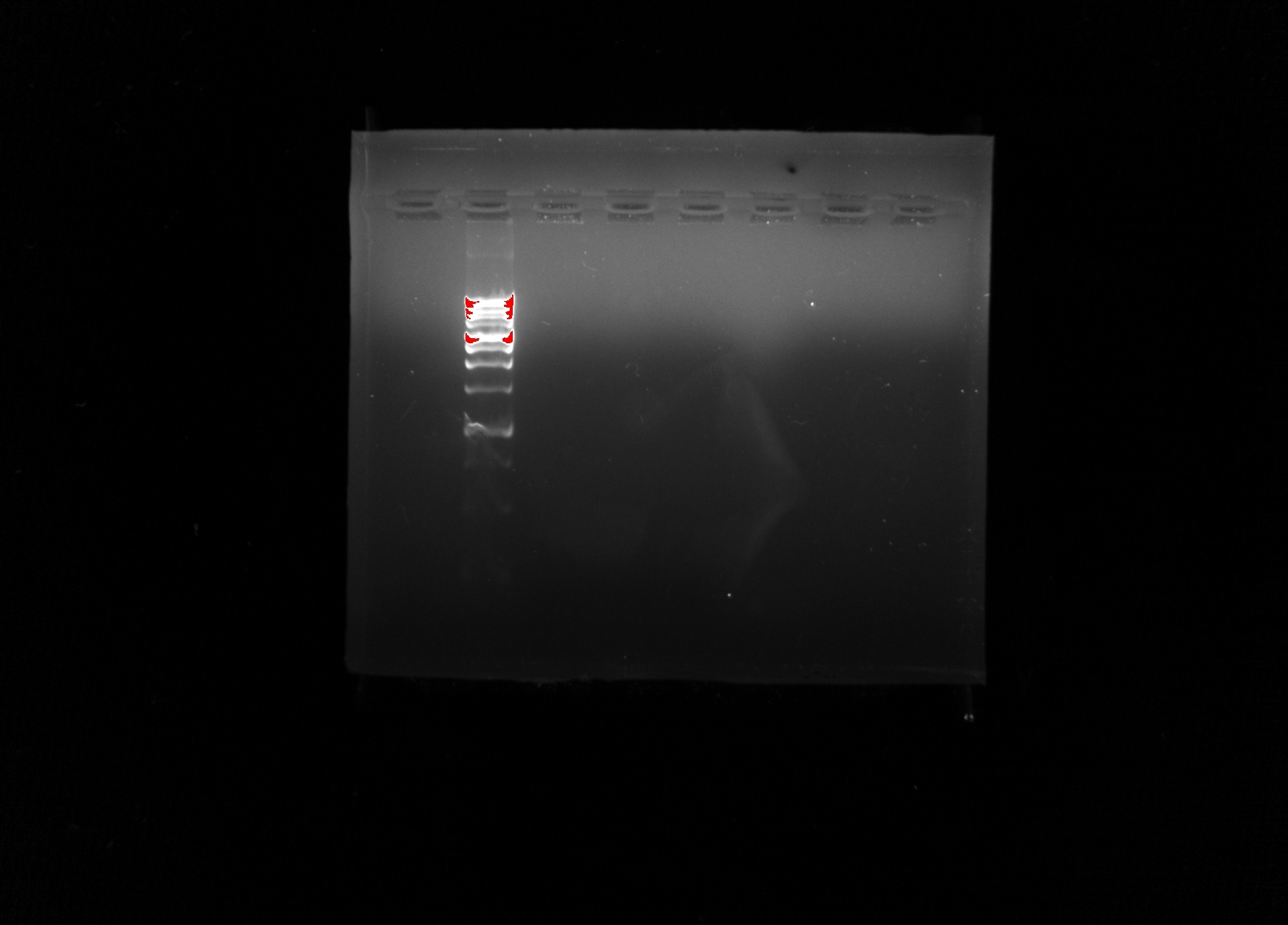
The image above depicts our gel after it was scanned by a gel imaging machine that used UV like to differentiate the bands from the gel itself. Our wells were used as follows: 1. Practice Loading Buffer One, 2. Ladder, 3. DNA sample, 4. Positive Control, 5. Negative Control, 6. Empty well, 7. Practice Loading Buffer Two, Practice Loading Buffer Three. Unfortunately, the only bands that are present after completion of gel electrophoresis. We can conclude that there was no DNA present in the samples that we had tested. Due to the fact that there were no bands present in the positive control then we can assume the cells that were present must have been destroyed in an earlier process. Without bands and fragment separation of base pairs, we are unable to continue the experiment and this step may have to be repeated with different DNA samples and positive controls.
Current Storage:
During lab we were unable to complete gel electrophoresis because the bands had only migrated half way through the gel after being in the gel boxes for thirty minutes at high power. After this, I assume that the gel way removed from the gel box and scanned with UV light under a gel imaging machine. These scans were uploaded to canvas as pdf’s that were labeled with each group’s number and course section number. My group’s pdf was labeled “group 5 -21”.
Future Goals:
In the future, I hope that we are able to see the fragments of DNA within the samples using gel electrophoresis. We may not be able to proceed with the metabarcoding process because we have no DNA to compare anything with. I hope that once we get a sample that will migrate down the gel and visually show the presence of bands/ base pairs, we may then be able to compare that with the DNA of other organisms that have the cox 1 gene to determine whether or not our DNA samples also contained that gene.