4/12/18

Purpose:
Today we completed gel electrophoresis on our samples that went through PCR last time. We also Imaged them using an ultraviolet imager. Lastly, we began to prepare our posters to present our findings.
Procedure:
- Remove the previously prepared 1.8% agarose gel from storage.
- Place the gel in the electrophoresis chamber and fill the chamber with 1x TAE until the gel is covered.
- Place 10 μl each of the samples into their own wells in the gel.
- Place 5 μl of the ladder in one of the wells
- Place the lid on the chamber.
- Connect the electrodes to the power supply unit.
- Run at 100v for 30 minutes
- Turn off the power supply and remove the gel.
- Image the gel with a UV imaging device.
Results :
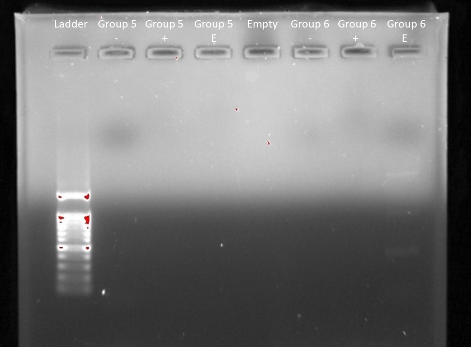
Our imaged agaros gel after electrophoresis. From left to right: Ladder, Group 5 Negative Control, Group 5 Positive Control, Group 5 Environmental Sample, Empty, Group 6 Negative Control, Group 6 Positive Control, Group 6 Environmental Sample.
Our group did not have an DNA show up in the imaging. However, group six had some show up in their experimental sample.
Conclusion:
I do not know why we had no positive results in our test. The only explanation I can think of, that would explain both the positive control and the experimental sample showing up negative, is that we did something wrong when preparing them for PCR. If the polymerase chain reaction did not work properly then nothing would show up during the imaging. However, I do not know what step we would have done incorrectly for both samples that would cause PCR not to work. I am especial curious about what went wrong, because our group used almost the exact same protocol for the previous test and we did have a positive result.