Lab 6: EZNA Tissue DNA Kit Protocol
2/15/18
Purpose: The purpose of today’s lab was to continue our lab from last week and use the protocol from the EZNA Tissue Kit to isolate ciliate DNA in our soil sample. This was our very first trial with the protocol, so it’s possible we may have to attempt again (but that’s totally okay!!). My group came in last Friday to isolate ciliates and take counts, so we were ahead. For the groups that didn’t come to lab last Friday, they had to isolate ciliates and take counts, so this resulted in some groups not getting as far as us. Today’s lab was really busy so it made time fly by.
Procedure:
A. Before starting
- Set the heat block to 70 degrees Celsius.
- Place the elution buffer into the heat block.
- Chill PBS on ice to cool to 4 degrees Celsius.
B. Prepare the cell suspension
- Was the cells with the cold 200 uL of PBS and vortex in the table vortex for 5 minutes then remove the supernatant.
- Resuspend the cells in 200 uL of fresh PBS.
- Add 25 uL of OB Protease solution and vortex for 5 minutes for a complete mix.
C. Lysis
- Add in 220 uL of BL buffer
- Incubate the tube for 5 minutes in the heat block and then vortex for a minute on the table vortex. Repeat this twice for a total of 10 minutes in the heat block and 2 minutes in the table vortex.
D. Binding
- Place 220 μl of 100% ethanol into the tube and vortex thoroughly for 1 minutes to mix completely
- Insert the HiBind DNA mini column into a 2 mL collection tube.
- Transfer the entire sample from step 2 above into the HiBind DNA mini column.
Note: The solution in my group was SUPER sticky and thick. We think this could be because our sample seemed to still have some soil and other debris.
E. Wash and Dry
- To fix this, we added 500 uL of binding HBC buffer into tube where our sample was rather than placing it in the column and then we vortexed our tube and column for 30 seconds at maximum speed (13,000 g).
- After doing this, our solution was still a bit too thick, we continued to add 500 uL of binding HBC buffer to dilute it down and vortexing again. This was done 3 times.
- After each spin we would discard the filtrate that came out of the solution after centrifugating and we’d use a kim wipe to clean our tube and use it again.
- We centrifuged one last time at maximum speed (130000) for 30 seconds. Then discarded the filtrate and the collection tube in the trash can, but kept the column.
- Place he HiBind DNA mini column into a new 2 ml collection tube.
- Add 700 uL of the DNA wash buffer to the column.
- Centrifuge the EMPTY HiBind DNA mini column at the maximum speed of 13000 g for 2 minutes to dry the column.
F. Elute
- Transfer the HiBind DNA mini column into a nuclease free 1.5 ml microcentrifuge tube and label with the name of the group and the date.
- Add in 100 μl of elution buffer heated to 70 degrees C.
- Then let the tube sit at room temperature for 2 minutes.
- Centrifuge the tube one last time at maximum speed at 13000 g for 1 minute so that the DNA is now floating in the solution.
- Store the eluted DNA sample at -20 degrees C in a refrigerator.
Note: Since Lillian and I were able to come to open lab, we were able to get ahead and complete the entire protocol today!
Results:
Last Friday, Lillian and I were able to come to open lab and this was SO helpful. In open lab we came to extract ciliates and get some counts. We took 3 2 uL drops on a concavity slide under a microscope and our counts were 21, 24, 25, 19, and 27. This resulted in an average of 23 ciliates per drop. Lillian did the math for our group and we concluded that there would be a concentration of 11500 ciliates per uL of sample. Most of the ciliates we seen were in cyst form. Some looked like they were clumped together, others were alone.
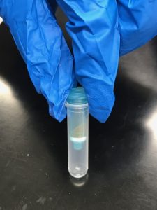
Storage: Our sample was stored in the orange microcentrifuge tube labeled LAK with 2/15/18 on the side and with a star on the top. Dr. Adair took our sample and placed it in a rack to store away in the refrigerator to cool at -20 degrees Celsius.
Conclusion: I had a fun time in lab. It made me feel good that my group and I were ahead! We sacrificed some of our Friday and it really paid off today in lab. I like the EZNA tissue DNA kit protocol. It seemed smooth and successful. Hopefully we did everything correct, if not there’s always time to revise this protocol to make it more efficient. I hope next lab were able to start PCR and amplifying our DNA!
Future steps: I’m so glad we have Dr. Adair in lab to help us out. Our group got stuck when our solution was heavily viscous. We had to improvise our protocol by thinking on our toes of what could help dilute the DNA. Hopefully this was a benefit to our DNA and were able to start PCR and amplifying DNA next lab, or at least learn about it in depth some more. The next step would be gel electrophoresis!! I’m so excited about that because I did a small experiment on it in high school and i’m stoked to revisit that.


