Lab 13: Identifying Ciliates 11/16/17

Objective:
The goal of today’s lab was to see if we were able to view any ciliates from the new culture of ciliates. The goal of today was to mainly observe whether or not we had successfully isolated ciliates from our non-flooded plates and if they had effectively been cultured. If we were not able to see any ciliates we had to go back to our non-flooded plates and repeat the process stated in lab 12.
Purpose:
The purpose of lab was to be able to view ciliates that we had isolated from our soil and possibly identify/ classify them. This would be helpful practice for identifying ciliates or even trying to identify some specific features of the ones that were able to be observed under the microscope.
Procedure:
1.) Retrieve the well plate with cultured ciliates.
2.) View the whole well under a dissecting microscope to see if any ciliates are present within the well.
3.) If ciliates were present within the well, pipet ten microliters onto a concavity slide and view under a compound microscope.
4.) If there were no ciliates present within the well, retrieve the non-flooded plate and try to isolate and extract a ciliate from that sample.
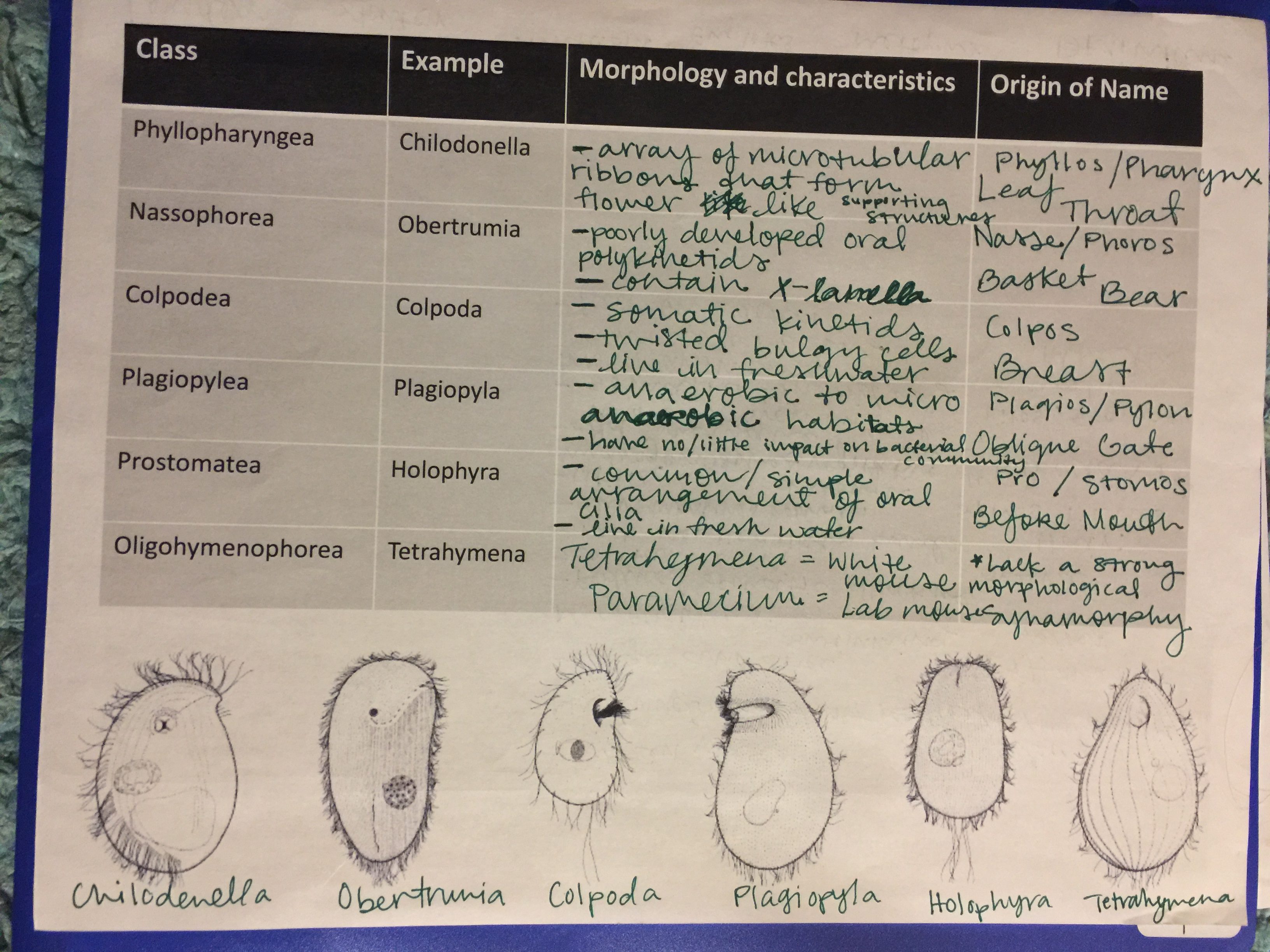
5.) Once a ciliate is able to be seen under a microscope, try to classify and and identify the ciliates present.
6.) Try to capture an image of any of the ciliates that can be seen under the micro scope.
Note: Some ciliates may be too small to identify using the compound microscope.
7.) These images should be used during your group presentation of your findings during this experiment.
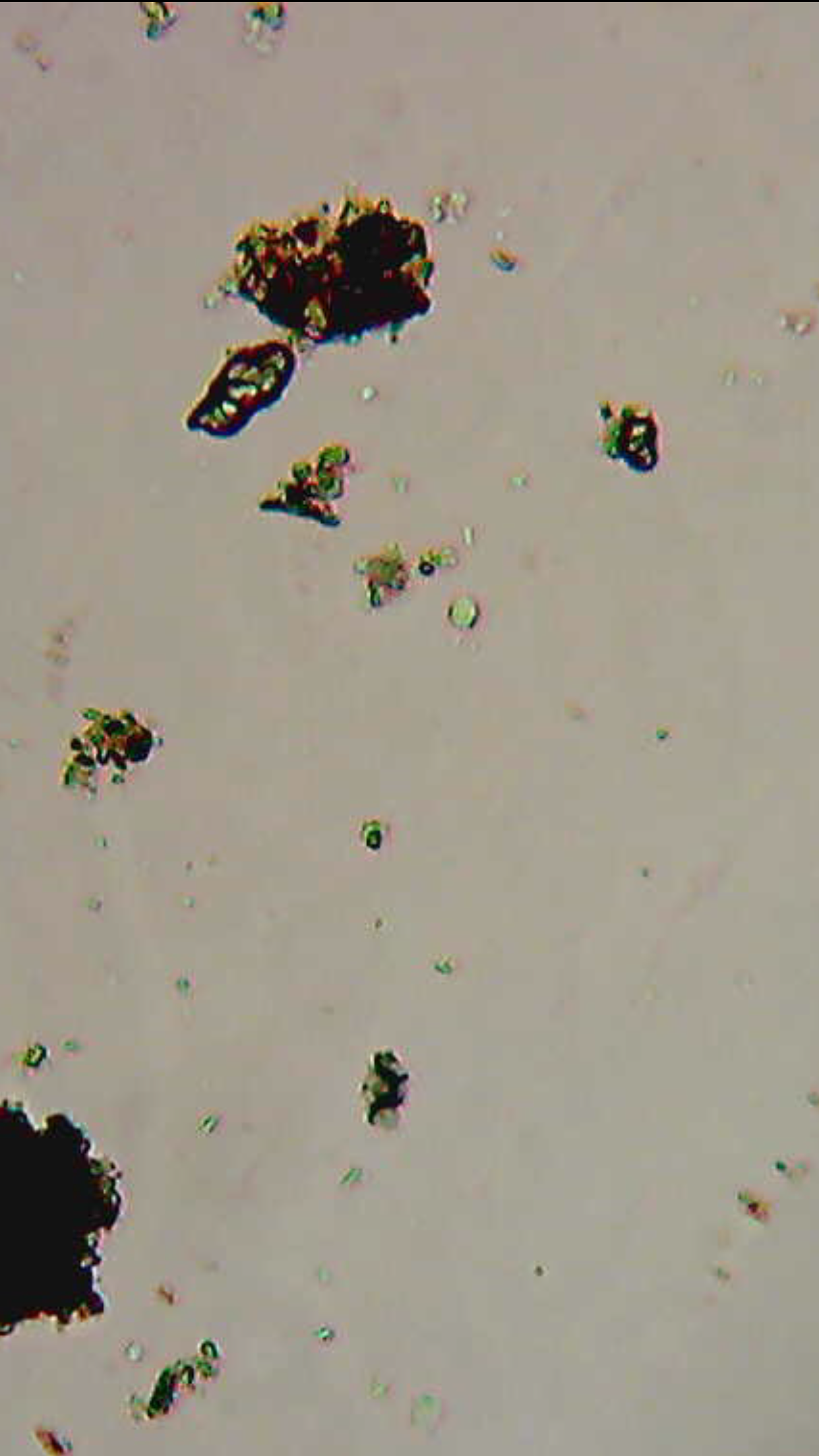
Data/Observations:
I observed a few ciliates that were too small to identify and could best be seen using 100x on the compound microscope. The ciliates I had seen were more oval shaped with very distinct organelles within due to its transparent body.


Current Storage:
My non-flooded plate and 24 well- plate are labeled with my soil identifier, KSA31F17, and are located in the left back desk in you are facing the projector in the second drawer on the front side of drawers closest to the next lab table. The falcon tube that was used to calculate the soil texture in located back in the teal- green tube stand with the other students tubes on the back lab counter closest to Dr. Adair desk top computer.
Future Goal:
I hope to be able to collect more soil samples in the future and be able to identify more ciliates or at least classify the group that the ciliate belongs to. I hope to use my knowledge I gained from this lab for any future research career that I choose to pursue.