Ciliate Isolation and Characterization 11/16

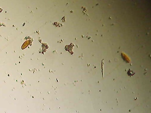
The Ciliate Isolation and Characterization lab was performed on 11/16. The purpose of this lab was to isolate, identify, and take pictures of the ciliates found in our soil. Since I was unable to find any ciliates in my personal sample of soil, I used the Burmuda grass sample. I took 0.5 µl of water from the Burmuda grass and tried to find ciliates under the compound microscope. When I was able to identify ciliates, I placed 0.5 µl of Methyl cellulose and 0.1 µl of iodine on the concave slide. This enabled me to capture a picture of the ciliates more easily and I also could see some of the internal structures a litter clearer. Next I used moticonnect to take a picture of the ciliates I found. Initially, I tried to use the coverslip so I could get a more detailed picture of my ciliate, but that failed. I had to re-do the procedure of finding ciliates from the Burmuda grass and the second time around, I did not use the coverslip. It is difficult to see a lot of the internal features based on the pictures I captured, however I was able to identify the macro and micronucleus before taking the picture. The ciliates are extremely small and they moved very quickly. Based on the picture, I would estimate that the dark ring around the ciliates are the cilia. It is difficult to identify the ciliate based on the picture alone, but my best guess would be that these ciliates are nassophorea’s. The next step would be to gather all of my information and present it to my classmates. I do not have a sample stored.