May 2

Lab 15: Poster Presentations

Taylor Hutcheson

Date:
April 28, 2017

Abstract
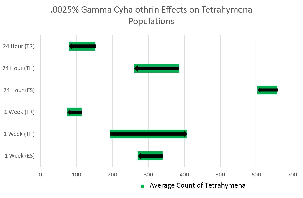
In our research, we gathered soil samples, picked out ciliates from these samples, and then extracted the DNA from the ciliates. Our objective was to extract DNA and use gel electrophoresis to determine the species that the DNA belongs to. Our gel electrophoresis results yielded no DNA, so our attempts to extract DNA were unsuccessful, therefore we were not able to identify a species based on DNA.

Introduction
Ciliates are microscopic single cellular eukaryotic organisms belonging to the phylum ciliophoran. They are characterized by their hair-like projections called cilia, which is where they get their name. Ciliates have two nuclei (one macro and one micro) and feed on small organisms such as bacteria through their oral grooves. They can reproduce sexually and asexually and play an important role in agriculture.

Materials/Methods

  1. Non-flooded Plates
    1. Add 10 g of soil and add water, place under microscope and pick out ciliates.
  2. Culturing
    1. Add picked ciliates to a medium of ceraphyll to encourage reproduction.
  3. DNA extraction
    1. Transfer 300 uL of ciliate culture to centrifuge.
    2. Incubate for 30 minutes in a 56 C water bath to denature proteins.
    3. Boil for 8 minutes and vortex for 1 minute.
    4. Centrifuge to pellet cellular debris
    5. Transfer supernatant, that contains DNA, to microcentrifuge tube.
  4. PCR and Gel Electrophoresis
    1. Denature proteins in a water bath of 94 C.
    2. 35 Cycles
    3. Denaturation: 94 C for 30 s
    4. Primer annealing 56 C for 20 s
    5. Primer elongation 72 C for 2.5 min
    6. Extension: 72 C for 5 Min
  5. Gel Electrophoresis =
    1. Prepare agarose gel with wells for DNA
    2. Run electrical to separate DNA fragments.
    3. Used as a tool to show genetic diversity (Jousset 2010)

Conclusion/Discussion
Because our gel electrophoresis results showed no bands or fragments of DNA, we came to the conclusion that we were not able to successfully extract the DNA from our ciliate cultures. This is also shown by the low amount of DNA in the results from the Nano Spectrophotometer. This could be be a result of not following procedures, technical difficulties, human error, or contamination. This research is significant because it proves “most ciliates are uncultivable and their population sizes are often too small, it is usually difficult to obtain sufficient genomic DNA required for PCR based experiments” (Kim 2009).

Literature
Jousset, A., Lara, E., Nikolausz, M., Harms, H., & Chatzinotas, A. (2010). Application of the denaturing gradient gel electrophoresis (DGGE) technique as an efficient diagnostic tool for ciliate communities in soil. Science of the Total Environment, 408(5), 1221-1225.

Kim, S., & Min, G. (2009). Optimization of DNA extraction from a single living ciliate for stable and repetitive PCR amplification. Animal Cells and Systems, 13(3), 351-356.

Shimano, S., Sambe, M., & Kasahara, Y. (2012). Application of nested PCR-DGGE (denaturing gradient gel electrophoresis) for the analysis of ciliate communities in soils. Microbes and Environments, 27(2), 136-141.

May 2

Lab 14: Gel Electrophoresis

Taylor Hutcheson

Date:
April 20, 2017

Disclaimer:
As does happen sometimes in scientific experiments, our PCR procedure did not go as planned. Our class learned that the machine that was supposed to hold our DNA samples at 4 C overnight malfunctioned, holding our samples at 94 C – a much higher temperature, resulting in the evaporation of all of our DNA. As a result, we (or rather, the TA’s) had to recreate the tubes used in last week’s experiment.

Goals:

  • Our objective was originally oriented on using our DNA after the PCR in a gel electrophoresis experiment so as to separate the DNA for identification, however, because this was no longer plausible, we focused on practicing gel electrophoresis until it was time to head to the computer lab and learn about how to create our posters.

Background:

Gel electrophorsesis separated fragments of DNA by size, with larger particles being closer to where they are placed and smaller particles traveling farther. The gel is placed in a salt-water solution so as to conduct electricity. The gel is stained by a DNA-binding dye and placed under a UV light, allowing the DNA to glow and become visible.

Procedure:

Gel Electrophoresis

  1. The TA’s recreated our treatment groups for us, this time not taking a control group into account due to limitations on time.
  2. After the TA’s recreated the mixture of Taq. Mix and EUK primers as according to last week’s procedure, we added the 5 uL of DNA solution, which we had preserved from our previous lab.
  3. We then vortexed the tubes for about 30 seconds, so as to ensure the reagents had mixed and settled to the bottom of the tube.
  4. The tubes were then sent again to another machine so as to run it through the process of heating and cooling (explained in last week’s lab).
  5. Finally, we practiced loading the gel electrophoresis trays with faculty DNA.

Computer Lab

  1. We reviewed how to create a proper scientific poster, learning that it is very important not to have too much text and to have many pictures.
  2. We then began brainstorming with our lab partners regarding how to go about the creation of our posters and would information we would like to include.

Data:

This is the photo of the results of my group’s gel electrophoresis after the TA’s ran our DNA through gel electrophoresis.

Future Experiments:
Because we our short on time, our next and final lab will simply include our poster presentations. If given more time, we would most likely try to redo the PCR and gel electrophoresis experiments with more pure concentrations of cultured DNA.

May 2

Lab 13: Posters and PCR

Taylor Hutcheson

Date:
April 13, 2017

Goals:

  • Prepare a PCR reaction using SSU ribosomal/universal primers (EUK). This will help to separate the cellular material from the nuclear material (though ribosomes may be an issue, as they have genetic material) in preparation for the gel electrophoresis.

Procedure:

  1. The control group will have an addition of 5 uL of water, while the treatment group will have the addition of 5 uL of DNA. Other than this, the process is the same for the two tubes. The control group is there to ensure that there were no outside factors or extra DNA found in the nuclear material.
  2. In the EUK tube, (mine labeled with a star), add 5.0 uL of DNA.
  3. In the control tube, add 5.0 uL of water.
  4. In both tubes, add 12.5 uL of Taq. Mix, 1 uL of EUK primers, and an additional 6.5 uL of water. After all this, the total volume of both tubes should be 25 uL.
  5. Vortex both tubes for a short amount of time to ensure that all of the reagents mix and settle to the bottom of the tubes.
  6. The tubes will now enter a thermal cycling profile.
    1. Initial denaturation: Heat the tubes at 94 C for 2.5 minutes.
    2. Now, the tubes will endure 35 cycles of:
      1. Denaturation: Heat the tubes at 94 C for 30 seconds.
      2. Primer annealing: Cool the tubes at 56 C for 20 seconds.
  • Primer elongation: Heat the tubes at 72 C for 2.5 minutes.
  1. Extension: Keep the tubes at 72 C for 5 minutes.
  1 EUK Tube Control Tube
2X Taq Mix (uL) 12.5 12.5
DNA (uL) 5.0 0
10 uM Euk Primers (uL) 1 1
Water (uL) 6.5 11.5
Total Volume: 25 uL 25uL


Future Experiments:
The next step will be to use the extracted DNA to perform gel electrophoresis, which uses an electrical current to distribute the nuclear material. From here, we will, in theory, be able to identify what type of ciliate the DNA belongs to by comparing it to the research of others.


May 2

Lab 12: DNA Extraction

Taylor Hutcheson

Date:
April 7, 2017

Goals:

  • Extract the DNA of dense ciliate culture so as to prepare for the process of gel electrophoresis.

Procedure:

  1. Transfer 300 uL – 500 uL of ciliate culture to microcentrifuge.
  2. Centrifuge for 5 minutes at 6000 g, discarding supernatant.
  3. Incubate for 30 minutes in a 56 C water bath, as this will break open the cells and denature some proteins.
  4. Boil for 8 minutes in 100 C water bath.
  5. Vortex for 1 minute.
  6. Centrifuge for 3 minutes at 16000 g to pellet cellular debris.
  7. Transfer supernatant with DNA in solution to microcentrifuge tube, without collecting the pellet.
  8. Label this tube with the soil identified (mine was TDH06S17) and place it in an ice box.

Observations:
After checking to see if my ciliates had cultured, I was pleasantly surprised to find that, under 40x magnification with a compound microscope, there were too many ciliates to count, all moving around quite rapidly.

Future Experiments:
After obtaining the DNA solution, the next step will be to prepare PCR reactions and gel electrophoresis so that we can identify the type if ciliate in my soil sample.

May 2

Lab 11: Characterizing Ciliates

Taylor Hutcheson

Date:
March 30, 2017

Goals:

  • Continue searching for ciliates within the non-flooded well plates with the intention of extracting ciliates from the solution and placing them in Cerophyll media so that they may culture.
  • The goal after obtaining cultured ciliates would be to extract their DNA so as to determine which type of ciliate the organism belongs to.

Procedure:
This lab is almost identical to the previous lab.

  1. Observe the non-flooded plate under both a dissecting and compound microscope, searching for ciliates to cultivate.
  2. When able to extract ciliates, use a pipette to extract about 2 uL of the ciliate and its surround solution, and place in a 24-well plate with 1000 uL of Cerophyll.

Observations:
After having no luck retrieving ciliates from the plate under a dissecting microscope due simply to how fast they swim, I decided to use a compound microscope. I took a small sample of water from the plate and placed it on a concavity slide, observing it at 4x, 10x, and 40x magnification. It was not until 40x that I found multiple small ciliates. I realized, after backing up back to 10x magnification, that I could observe the ciliates causing small solid particles such as dirt and shrubbery to move with their motion through the solution. I then followed the procedure and placed multiple of these ciliates in well A1 of my 24-well plate with the Cerophyll media.

Future experiments:
If, by next week my ciliates have cultured, I will be able to move on to the DNA extraction of the dense ciliate culture.

May 2

Lab 10: Soil and Ciliates

Taylor Hutcheson

Date:
March 23, 2017

Goals:

  • Complete testing and analysis for the metadata of the soil sample.
  • Continue the process of extracting ciliates from the non-flooded plate.

Procedure:

  1. Use drops of water from the non-flooded plates to determine the pH of the soil.
    1. Using a plastic pipette, drop some of the water from the plate onto a piece of pH paper.
    2. Compare the color on the paper to that of a key.
  2. Weigh the dry soil sample and compare it to its weight last week.
  3. Continue searching soil samples for ciliates.

Observations:
After allowing my soil to sit in the non-flooded well for a week, I had high hopes that the ciliates would come out. Towards the beginning of the class period, I did not find a single organism. About an hour in, I finally found a very small, circular ciliate moving very quickly through the water-soil solution. I would then find one about every 30 minutes after that, but always moing too quikly to pluck from the solution using the pipette. I found these ciliates in the center of the plate, always near a body of soil rather then in an ocean of water (or what would be an ocean to a ciliate). Once, on the outer edge of the plate, I found a relatively larger and longer ciliate. I still good not harvest a single ciliate. I even tried taking small droplets of about 400 uL of the non-flooded plate’s water, and analyzing just those droplets.

Data:

  1. pH: 6.0 – slightly acidic
  2. Soil type: Silt loam
    1. Silt 58.8%
    2. Sand 29.4%
    3. Clay 11.8%
  3. Dry weight of soil: 4.2 grams
    1. Water made up 0.8 grams of the soil’s weight

Future Experiments:
From here, it will be beneficial to continue the search for ciliates. I think a strategy I will pick up next lab will be checking a sample of soil under a compound microscope.


May 1

Lab 9: Soil Collection

Taylor Hutcheson

Date:
March 16, 2017

Goals:

  • Investigate the different types of ciliates and other microorganisms living in the soil samples we collected during our Spring Break.
  • Become familiar with the practice of using non-flooded plates and observing them under a dissecting microscope, as well as becoming familiar with what to look for and how to identify ciliates.

Background:
Prior to Spring Break, our lab was directed to collect at least one sample of soil from our Spring Break destination with the goal of identifying a variety of small organisms – primarily, ciliates. I obtained my own soil from grass on the side of a sidewalk in the National Mall in Washington D.C., an extremely and constantly populated area. I wondered how this would affect the diversity in my sample, hoping the constant human movement would not disturb the organisms I would find. Other members of my group found their soil samples in New Orleans, LA; Jacksonville, FL; and London, England.

Procedure:
Non-flooded Plate

  1. Place approximately 10 grams of air dried soil in a dish.
  2. Saturate the soil with water and give time for the soil to soak.
  3. Observe the soil under a dissecting microscope and look for ciliates.
  4. Keep micropipettes ready so as to pluck any found ciliates from soil. From there, one would place the ciliate in a 24-well plate with cerophyll media, so as to allow it to culture.

Soil Metadata

  1. First, prepare a test to analyze water content in the soil.
    1. After weighing and taring a boat weighing the soil, add approximately 5 grams of wet soil into the boat and record the total mass.
    2. This soil will be weighed again in a week.
  2. To determine the type of soil in the sample, prepare a soil identifier test.
    1. Put about 5 mL of soil into a falcon tube with your soil identifier on it. My soil identifier was TDHS06S17.
    2. Add D.I. water into the falcon tube until the water reaches the 10 mL mark.
    3. Add 1 drop of suspension solution to the mixture of water and soil, them vortex the tube until the soil and water are thoroughly mixed.
    4. Put the falcon tube in a tray and allow to settle for a week.

Observations

Non-flooded Plate
My soil sample was a rich, dark brown color and seemed relatively high in water concentration. When I first opened my soil bag, a small spider sprang out. My soil was also very rich in shrubbery, as it had been collected from a grassy area under a tree.

Soil Metadata
To be determined next week.

Future Experiments
Next week, I will need to continue obtaining the metadata on my soil, completing the water content test and the soil identifier test, as well as testing the soil pH. In the coming weeks, we will continue to observe our soil using both a dissecting and compound microscope. The ultimate goal is to pluck ciliates from the soil and allow them to culture, and eventually obtain the ciliate’s DNA.

May 1

Lab 8: Peer Review and Soil Ciliates

Taylor Hutcheson

Date:
March 9, 2017

Goals:

  • Read and revise the lab reports of our peers so as to aid them and recognize how our own lab reports should look.
  • Explore the concept of the significance of soil biodiversity and its role in our environment.
  • Practice setting up a non-flooded plate well plate with previously used soil so as to prepare and become familiar with the practice we will use after obtaining our own soil samples.

Procedure:
Peer Revisions

  1. Trade papers so that no one who tested the same concentration of Tetrahymena revised each other’s papers.
  2. Revise papers according to the rubric, while making annotations and comments on the paper.
  3. List of questions to grade the paper by:
    1. Is the title descriptive?
    2. Are all of the authors included?
    3. Does the abstract have the Background, Purpose, Methods, Results and Conclusion?
    4. Is it 250 words or less?
    5. Is the introduction formatted correctly, moving from general to specific?
    6. Does the introduction have at least 3 references from the primary literature?
    7. Are the methods written in paragraph form using the passive voice?
    8. Do the methods parallel the results?
    9. Are the methods clearly written so that others could repeat the experiment?
    10. Are the results represented in paragraph form and in appropriate figures and/or tables?
    11. Are the figures labeled, numbered, and include a caption?
    12. Is there an effort to interpret and analyze the results in the discussion?
    13. Are any conclusions formed?

Non-flooded Well Plate

  1. Obtain a plate and a soil sample.
  2. Add about 10 grams of soil onto the plate.
  3. Using a plastic pipette, add enough D.I. water to cover, but not flood the plate so that the soil is saturated.
  4. Observe the non-flooded plate under a dissecting microscope with the main goal of observing ciliate organisms.

Observations:
I did not observe any ciliates in the soil of my own non-flooded plate. However, I was able to observe two microscopic tick-like creatures, both attached to a small piece of wood in seemingly random locations in my soil. I was also able to observe a worm-like creature, which moved its way slowly through the soil. This small worm creature was most likely a nematode.

Future Experiments:
In the future, we will collect samples of our own soil in whatever location we visit during our Spring Break. Because all the members of my group are heading to very diverse locations around the United States, and even internationally, observing the differences in our soil samples will hopefully give our group a bit of variety in observed organisms and types of ciliates. In the future, we will also be completing our lab reports.

February 27

Lab 7: Results and Analysis

Taylor Hutcheson

Date:
23 February 2017

Title:
Results and Analysis

Goals:

  • Collaborate with your group members so as to come up with the most effective way to visualize and portray the data you have all recorded.
  • Learn how to activate and use the data analysis tool pack in Microsoft Excel.

Procedures:

  1. Observe other presentations and assist in the critique of their graphs.
  2. Present our own group’s graph.
  3. Enable the data analysis toolpack in Microsoft Excel.
  4. Use this toolpack to generate desired statistics about selected groups of data regarding the cell counts of Tetrahymena cultures.
  5. Use this data to create bins.

Observations & Data Analysis:

It was noted during this meeting that a line graph would not be appropriate for our range of data as it traditionally has to do with change over time, whereas the data we have collected does not have a constant change. This is important to keep in mind in the future.

Our group initially planned on doing a bar graph, however, with the large range of data collected between the three of us, we found this method to be too cluttered to fit onto one graph. This appears to have been a problem with other groups, as well, as those who decided on the bar graph ended up with many bar graphs.

Though the graph our group came up with ended up something somewhat out of the box, it is important to keep in mind when moving forward that, as was mentioned in class, “there is always a better way to graph it.”

Next Steps:
The next steps will include the implementation of professional data analysis, especially when it comes to writing scientific papers regarding our research.


February 27

Lab 6: Evaluating the Experiment

Taylor Hutcheson

Date:
17 February 2017

Title:
Evaluating the Experiment

Goals:

  • Complete the one week cells counts and serial dilutions we began one week prior to this lab.
  • Become familiar with the use of Methyl green-pytonin staining using a wet mount.
  • Compare date between the 24-hour cell counts and the 1-week cell counts, as well as its relation to cell groups that were and were not treated with pesticide.

Procedures:

  1. Retreive the cultures and observe under a dissecting microscope to see if any cells are still alive.
  2. Transfer 2 uL from the control and treatment wells to a concavity slide and observe under a compound microscope so as to count the number of living cells in each.
  3. Dilutions may be necessary if there are too many cells to count.

Observations & Data Analysis:

  • C1 – control, 24 hour – 285 cells
  • C2 – control, 24 hour – 145 cells
  • C3 – control, 24 hour – 35 cells
  • C4 – control, 7 days – 160 cells
  • C5 – control, 7 days – 265 cells
  • C6 – control, 7 days – 155 cells
  • D1 – treatment, 24 hour – 105 cells
  • D2 – treatment, 24 hour – 160 cells
  • D3 – treatment, 24 hour – 1115 cells
  • D4 – treatment, 7 days – 145 cells
  • D5 – treatment, 7 days – 315 cells
  • D6 – treatment, 7 days – 180 cells

Next Steps:
The next steps would be to create a graph so as to portray all of the data from my group.