January
27
Lab 2: Meet the Ciliates
Date: 1/26/2017
Title: Lab 2: Meet the Ciliates
Goal: Observe, identify, and characterize ciliates according to shape, size, movement, and location using a dissecting microscope.
Background: We researched proper lab procedures and studied laboratory safety. We researched ciliates and their importance.
Procedures:
- Clean desk with 10% bleach solution in spray bottles
- Obtain a clean 24-well plate
- Using the provided plastic pipette, place each unknown into separate well. *Note: Document which unknown you placed in which well. Fill wells about half full. **NOTE: it is important to keep the unknown cultures pure- do not use the same plastic pipette on more than one unknown! Only use the pipettes provided with each culture
- Observe each unknown under the dissecting microscope and fill out the table to the best of your ability.
- Make a detailed sketch of each ciliate, and provide a tentative identification along with your reasoning.
- Tape your observations and notes into your lab notebook and show your work to an instructor.
- Discuss the “Questions That Matter” with your group, write your answers, and turn in to your T.A.
Observations:
| Unknown # | Shape | Relative size | Movement | Location in Media | Other characteristics |
| E in A1 | -trumpet shaped
-sometimes bell-shaped -spherical bodies |
150-3000 microliters | -free swimming
-spiral movement |
surrounding detritus or some organic matter | green |
| D in A2 | – oval shaped | 30-220 microliters | -free swimming, spiraling | all over the well | very small |
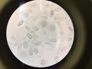
| C in A3 | – elongated, oval shaped, ovoid | 60-300 microliters | -free-swimming | all over the well | light purple color |
| B in A4 | -elongated, no spine | 200-300 microliters | -free-swimming, back and forth movement, circular movement | along the rim of the well | very thin and worm-like |
| A in A5 | -elongated, twisted, no spine | 30-300 microliters | -free-swimming, spiral movement | all over the well | thin |
Data Analysis:
- Unknown E in A1: Stentor
- Unknown D in A2: Euplotes
- Unknown C in A3: Blepharisma
- Unknown B in A4: Spirostomum
- Unknown A in A5: Paramecium
Next steps: Read the lab procedure more carefully next time. Learn more about ciliate behavior and movement.