August 29

Phage Annotation #1 8-29-17


  • Background
    • What is gene annotation?
      • Using the raw DNA sequence information, and studying the sequence looking for structural and functional aspects of the genome. Picking relative locations of genes in a genomic sequence including protein- encoding genes and RNA encoding genes
      • Studying the genome of various DNA sequences gives the scientific community information on both the biological and biochemical functions and roles the DNA plays within organisms and within biological groups.
  • Objective
    • To present and discuss recent scholarly articles regarding research on phages and health
    • To become familiar with the processes of uploading and annotating a gene
    • To make predictions of the function of a gene or region of a gene using DNA master
  • Procedure
    • We first set key preferences in DNA master by selecting File/ Preferences/ Local Settings/ Colors, and set the display colors for the tRNAs, tmRNAs, and ORFs. Next, we clicked local settings/ codons, and checked “Use TTG start codons” and “apply”
    • Next we imported a DNA sequence from the phagesdb database called, “Amigo”. Due to technical difficulties on DNA master, we uploaded an already finalized “Final DNA Master File” by selecting file/ open/ DNA Master file/ open
    • Next we annotated the genome by selecting genome/ annotation/ Auto-annotate
    • Next we refined our positional annotation features by selecting features and choosing codon #24, and under the notes section we listed, “SSC: start 11810 stop 12712 gap: 902”.
    • From the DNA master menu, we selected DNA/ Frames/ ORF Analyis
    • We compared potential start codons by selecting frames/ ORF/ 5’-3’ / Choose ORF Start. This sequence listed each possible start codon.
  • Results

Figure 1: Possible Start Codons for Gene #24 Phage: Amigo

  • Figure 1 is a table listing the possible start codons for gene #24/ the selected ORF window, the upstream nucleotide sequence, and the gene length
  • Conclusion
    • The results of this lab are the first pieces of information necessary for performing a detailed and refined annotation. Although this gene has already been annotated and submitted, future annotations will teach us information about the structure and function of a given gene, which has potential benefits to the scientific and health community.
  • Future Work
    • Successive steps to be taken for this lab incred creating a spreadsheet of current gene calls, and creating an ORF map of current gene calls. Running a gene blast on this particular gene can give us information on protein sequences that could provide more information on the gene itself.
    • Running more and more annotations will allow the class to become more familiar with DNA Master as a program, and thus will build a knowledge base for students to interpret the significance of patterns and aspects of the nucleotide sequence itself.
May 4

Final Summary

  • I used two samples for soil collection, therefore I had two soil identifiers. The numbers appearing after the dash indicate how far away from the shore the soil was collected.
  • The two identifiers I used were TRR06S17-50 and TRR06S17-2
  • To get the soil, I used a shot glass and scooped up the first 2-3 inches of soil.

Soil Metadata for TRR06S17-2 and TRR06S17-50

Coordinates: 30.3028196, -89.7928000

Date Sampled: March 12, 2017

Time Sampled 2:00 PM

Temperature: 62° F

Humidity:85

Last Rainfall: March 11, 2017


TRR06S17-2

Soil type: sandy loam

  • 4.88% Clay
  • 46.3% Silt
  • 48.8% Sand

pH: 6

Percent Water: 17.65%

TRR06S17-50

Soil type: loamy sand

  • 0% Clay
  • 39% Silt
  • 61% Sand

pH: 6

Percent Water: 17.65%

Collection

Once we obtained our soil samples, we had to collect some initial metadata, such as the percent water, pH, and the different components of the soil. To collect the percent water, we weighed out ~5 grams, and then let it sit out for a week. The water evaporated, and then the new weight was the “dry weight”. To get the percentages of the different components, we added water and a drop of resuspension solution to separate the different layers. And lastly, to get the pH, we used pH test paper, and then compared the color to a chart of different pH values and colors.

Non-flooded Plates

After collecting this metadata, the hunt for ciliates began. We began by creating non-flooded plates. These are created by placing a small amount of soil into a dish, and then adding DI water until some of it runs off. In this water run off is where the majority of ciliates will be found. It is important to not over water the samples, hence the term “non-flooded”. We mainly used a dissecting microscope, but occasionally we would put samples of the water onto concavity slides to look for smaller ciliates. Any ciliates that were found were placed into a 24 well plate with ~1000 uL of Ceraphyll, a grass extract which promotes ciliate reproduction. Because ciliates can reproduce asexually, a single ciliate can eventually become an entire culture.

Confirming DNA Presence

I was not able to find any ciliates in my first two non-flooded plates, so after three unsuccessful weeks, in an open lab I created two new non-flooded plates. However, even though I found lots of ciliates, none of them would create successful cultures. Because of this, the TA in the open lab suggested that I just pick as many as I could, which ended up being like 20, and then attempted DNA extraction. Below is my nanodrop results from the open lab with my “culture” of 20 ciliates. The process of extraction involved transferring 300 uL of ciliate culture to a centrifuge, and then incubating that in a 56° C water bath in order to denature the proteins. After the proteins were denatured, the samples were boiled and then vortexed. Next, the samples were centrifuge in order to collect all the cellular debris in a pellet. Then, the supernatant, the liquid on top,  was transferred to a micro-centrifuge tube. One drop of this was placed on a nano spectrophotometer to assess the amount of DNA. The label I used on my centrifuge tube for my DNA was TRR06S17-DNA.

Polymerase Chain Reaction

Using the DNA in our micro-centrifuge tubes, we amplified the DNA by the process of PCR, which stands for polymerase chain reaction. The process of PCR involved denaturing the proteins holding the double helix together, then applying primers and TAQ Polymerase to replicate the strands. We use TAQ polymerase because it is resistant to the heat changes that denature the proteins. This process is repeated many times to create multiple copies of the same sequence. The tube we created for PCR had a certain number and letter assigned to it in order to keep track of whose sample was whose, my tube was labeled G6.

Gel Electrophoresis

After PCR was repeated many times, the amplified samples were loaded into gel electrophoresis wells which were prepared with agarose solutions. The DNA is loaded into slots in the agarose gel, and then an electric current is ran through the gel and DNA, and because of the negative charge of DNA, the strands migrate toward the positive end of the circuit. The shorter fragments move farther faster, and this creates a “ladder” or “barcode” that can be used to identify the species where the DNA came from. My results from gel electrophoresis showed no DNA, therefore I made an error while loading the DNA, extracting the DNA, or amplifying the DNA.

Conclusion

In conclusion, while the experience was great, I yielded no DNA results. This is not to say that the entire lab was a failure however, because I became acquainted with many lab techniques, and I am sure if I was to repeat the process, I would do it much more efficiently. Science is not always about finding some new breakthrough idea, science is also the all the failures that came before that breakthrough moment. In the future, I look forward to being a part of the 1106 research lab, and I am sure that the skills I learned in this lab will be invaluable. Still, further ciliate research needs to be done, because they are one of the important aspects of healthy soil, and if we are trying to deal with an ever-growing population and a food-scarcity, ciliates might just be the answer that we need to feed this world. Because of this, more research needs to be done on how ciliates can affect soil health and by extension crop yields.


May 3

final summary

Soil collection:

My soil sample: label as JJ06S17

GPS Coordinates: _31*33’32” N, 97*7’27” W

Location: By the Brazos River, 3 foot from the river water, probably just river mud

Environmental Conditions

  • Date sampled : __3/10/2017__________
  • Time of day : __5:30pm______________
  • Temperature : __73/63F_______________
  • Humidity : ____81%___________________
  • Last rainfall : ___3/7/2017______________

Set up non-flooded plate

Protocol:

  1. put 10-50 g of fresh or air-dried soil in a petri dish (label as JJ06S17)
  2. Saturate but do not flood the sample with DI water. Add water until about 5-20 ml will drain off when the petri dish is tilted. Wait for a little bit time.
  3. Observe your soil using the dissecting microscope and record observation.

After 1-7 days

  1. Observe them and try to separate individual one


Soil metadata

  • set up water content test: weight the weigh boat; add~5 grams of soil; record mass of wet soil
  • Label 1 falcon tube with my soil identifier (label as JJ06S17). Add soil (remove plants. sticks and large clods) to the 5 ml mark and add water to the 10 ml mark. add 1 drop of the responsion solution and mix well
  • let the tube sand for 1 minute and then pour off the suspension into a second falcon tube
  • What remains in my first tube is sand. record the volume of sand
  • calculate the  % sand in my soil
  • label the second tube with the soil identifier and save for next week
  • Take a drop of the water from non-flooded plate on a pH paper. After color changes,


Water content calculation: (fresh wet soil – dry soil)/fresh wet soil = 21%

Soil characteristic: % sand, silt, and clay

1) Sand=92% 2) silt=0.4% 3) clay=8% which means my soil sample identified as sand

pH= 6.5


Discovering new ciliate


Observe from the well plate and see if there are any ciliate inside and try to separate one and culture for a large number for further study.

Pick, culture, and characterize

From last week of the non-flooded plate, I try to separate the ciliate

First: observe to find one

Observe the non-flooded plate under the dissecting scope

Using micropipette to get few microliters of the water from the edge of the non-flooded plate

Put it one a concavity slide and observe under a compound scope

Based on unmoved sand or other references, you can see some small ciliate moving.

Second: isolated one ciliate

Using serial dilution to separate one

Be careful to using a tip to collecting them

Third: culture

Put them in to a well have enough PPT media

Wait for a week for them to growth

Characterize

After a week with enough food supply, they might grow into a large number

Take some to observe under the compound scope

Under the lower power, you can see their general size in a big way

Under the higher power, you can observe their movement and probably inner organization

Observation:

I separate my soil ciliate from my soil sample (labeled as JJ06S17 -2)

Try to separate one of them and cultural them for a week, my ciliate reproduced into hundreds of them.  (From well B3)

I think my ciliate was tetrahymena.

We need much more than the number I got, then I try to culture much more of them

I put 2 uL of the ciliate sample from my well B3 in to each well of the C row, and adding 1000 uL of the PPT media inside.

After another week of culturing them, I got most hundreds of in each well of the C row

The teacher said the PPT might be contaminated. I am afraid that my sample got contaminated. Therefore, I took some of the contaminated PPT media observe under the microscope compare with the ciliate I got. Luckily, most of them cooks different. In this way, I know my sample was not contaminated.


DNA distraction:

Procedure:

DNA extraction

Modified Chelex Extraction adapted from Stüder-Kypke, 2011.

  1. Transfer 1mL dense ciliate culture (20 or more individuals) to microcentrifuge tube.

– Make sure to record which ciliate culture you are extracting from

– Label your tube with your soil identifier

  1. Centrifuge @6000g for 5 minutes, discard supernatant
  2. Add 200uL 5% Chelex 100 to pellet, and vortex for 1 minute

– For this step, use large-bore micropipette tips or simply cut off the tip of a 1000uL micropipette tip

  1. Incubate for 30 minutes in 56oC water bath
  2. Boil for 8 minutes in 100oC water bath
  3. Vortex for 1 minute
  4. Centrifuge @ 16000g for 3 minutes to pellet cellular debris and Chelex beads
  5. Transfer supernatant to clean microcentrifuge tube, being careful not to transfer Chelexbeads
  6. Carefully label top and side of microcentrifuge tube with your soil identifier (e.g.TLA09F16A), date (MMDDYY = 062116), the well culture was removed from (e.g. B1 or D4), and “Chelex”


I label my tube as JJ06S17 from well C4.

After follow the procedure of DNA extraction, we did nanodrop test during the open lab.

The result as follow.

61 ng/uL

Ratio: 1.36


PCR

The polymerase chain reaction (PCR) is a technique used in molecular biology to amplify a single copy or a few copies of a piece of DNA across several orders of magnitude, generating thousands to millions of copies of a particular DNA sequence. It is an easy and cheap tool to amplify a focused segment of DNA, useful in the diagnosis and monitoring of genetic diseases, identification of criminals (under the field of forensics), studying the function of targeted segment, etc.


Procedure:

using SSU ribosomal/ universal primers(EUK).

Tube 1 (EUK) 3(control group)
2X Taq Mix (uL) 12.5 12.5
DNA (uL) 5.0 0
10uM EUK primers (uL) 1 1
Water uL
Total volume (uL) 25 25
  1. Perform PCR assay in 25 uL of reaction mixture

5 uL 2X Master Mix

5uL of your ciliate’s DNA

0 uL of the 10 uM primers to the tubes

(1.0uL)*(10uL)=X(25uL)

Final concentration = 0.4uL solution of each primer

Calculate the amount of water required for each tube o make the total volume 25 uL

Add uL water to equal to 25 uL

For each set of reagents, you need to run 1 control tube, the control will have extra water in place of the DNA

Mix your samples briefly by flicking the tube or vortexing. Give the tubes a quick spin to make sure all the reagents are in the bottom of the tube

  1. thermal cycling profile: follow the instructions to program the thermocycler for the following conditions

35 cycles:

Denaturation: 95° C for 30 s

Primer annealing: 56° C for 20 s

Primer elongation: 72° for 2 min and 30 s

Extension: 72° C for 5 min


Gel Electrophoresis Protocol

Part 1 Making an Agarose Gel (We did this last week)

**For this lab, you should be wearing latex gloves for the entire class period! Ethidium bromide is a known mutagen that intercalates between the bases in the DNA – great for identifying DNA on our gels, not so great for the cells in our bodies**

  1. Make 1xTAE (Tris acetate EDTA) in 1L Erlenmeyer flask from stock solution

For 1 Liter of 1xTAE, I will need _________ mL of the _____xTAE stock solution and _________ mL of D.I. water

  1. Make 1.5% agarose gel
    1. Add 40 mL 1xTAE to ______ g agarose in small Erlenmeyer flask
    2. Cover lightly with weighing paper/Kimwipe and loose-fitting cap (do NOT tightly close the container, this is how things end up exploding!)
    3. Heat until solution is clear and small bubbles come off the bottom when gently swirled
    4. Allow to cool (5-6 minutes)
    5. Add 2 μL ethidium bromide, swirl gently
  2. Set up gel electrophoresis box, making sure the open ends are somehow sealed (with tape, turned sideways into box, ect), and that the comb is inserted with its back towards the nearest edge
  3. Pour agarose gel smoothly into prepared mold, with as few bubbles as possible. Allow to sit at least 30 minutes to solidify
  4. Cover gel with prepared 1xTAE buffer solution so that it will not dry out
  5. Carefully remove comb and turn gel so that the wells are furthest away from positive electrode (think “run to red” – you want the DNA to run towards the red electrode)

Running the Agarose Gel

  1. Using a micropipetter, add 5 µL of the ladder and 10 µL of each PCR product. If the loading buffer is not included in the Taq polymerase used in the PCR, add 5 µL 5x loading buffer to the 25 µl PCR reaction and mix thoroughly before transferring 10 µl to the gel.
  2. After you have loaded your samples, place the lid on your box and turn on the power supply to approximately 100 volts. Allow to run for 30 minutes or more, allowing the loading dye to run approximately halfway across the gel before turning off the power.
  3. Image with UV light.


Result:

The two small tube for PCR test labeled as 6J and another control group as 6JC

During the whole process, we have to wearing gloves make sure the table was clean from contamination.


poster :

summary:

during this process, I’m better understand the way to do research. I’m super exited to find those ciliate inside my soil sample. Ciliate are wildly found but really hard to seperate. working in a group is hard. making a poster is hard. doing a presentation is hard. but the process is fun. Never give up

May 2

Lab 15: Poster Presentations

April 28, 2017

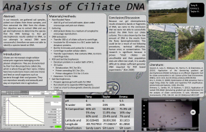
Analysis of Ciliate DNA

By: Tony R., Emma S., Taylor H.


Abstract:

In our research, we gathered soil samples, picked out ciliates from these samples, and then extracted the DNA from the ciliates. Our objective was to extract DNA and use gel electrophoresis to determine the species that the DNA belongs to. Our gel electrophoresis results yielded no DNA, so our attempts to extract DNA were unsuccessful, therefore we were not able to identify a species based on DNA.

Introduction:

Ciliates are microscopic single cellular eukaryotic organisms belonging to the phylum ciliophoran. They are characterized by their hair-like projections called cilia, which is where they get their name. Ciliates have two nuclei (one macro and one micro) and feed on small organisms such as bacteria through their oral grooves. They can reproduce sexually and asexually and play an important role in agriculture.

Materials/Methods:

  1. Non-flooded Plates
    1. Add 10 g of soil and add water, place under microscope and pick out ciliates.
  2. Culturing
    1. Add picked ciliates to a medium of Ceraphyll to encourage reproduction.
  3. DNA extraction
    1. Transfer 300 uL of ciliate culture to centrifuge.
    2. Incubate for 30 minutes in a 56 C water bath to denature proteins.
    3. Boil for 8 minutes and vortex for 1 minute.
    4. Centrifuge to pellet cellular debris
    5. Transfer supernatant, that contains DNA, to microcentrifuge tube.
  4. PCR and Gel Electrophoresis
    1. Denature proteins in a water bath of 94 C.
    2. 35 Cycles
    3. Denaturation: 94 C for 30 s
    4. Primer annealing 56 C for 20 s
    5. Primer elongation 72 C for 2.5 min
    6. Extension: 72 C for 5 Min
  5. Gel Electrophoresis
    1. Prepare agarose gel with wells for DNA
    2. Run electrical to separate DNA fragments.
    3. Used as a tool to show genetic diversity (Jousset 2010)

Conclusion/Discussion:

Because our gel electrophoresis results showed no bands or fragments of DNA, we came to the conclusion that we were not able to successfully extract the DNA from our ciliate cultures. This is also shown by the low amount of DNA in the results from the Nano Spectrophotometer. This could be a result of not following procedures, technical difficulties, human error, or contamination. This research is significant because it proves “most ciliates are uncultivable and their population sizes are often too small, it is usually difficult to obtain sufficient genomic DNA required for PCR based experiments” (Kim 2009).

Literature:

Jousset, A., Lara, E., Nikolausz, M., Harms, H., & Chatzinotas, A. (2010). Application of the denaturing gradient gel electrophoresis (DGGE) technique as an efficient diagnostic tool for ciliate communities in soil. Science of the Total Environment, 408(5), 1221-1225.

Kim, S., & Min, G. (2009). Optimization of DNA extraction from a single living ciliate for stable and repetitive PCR amplification. Animal Cells and Systems, 13(3), 351-356.

Shimano, S., Sambe, M., & Kasahara, Y. (2012). Application of nested PCR-DGGE (denaturing gradient gel electrophoresis) for the analysis of ciliate communities in soils. Microbes and Environments, 27(2), 136-141.


May 2

Lab 15: Poster Presentations

Taylor Hutcheson

Date:
April 28, 2017

Abstract
In our research, we gathered soil samples, picked out ciliates from these samples, and then extracted the DNA from the ciliates. Our objective was to extract DNA and use gel electrophoresis to determine the species that the DNA belongs to. Our gel electrophoresis results yielded no DNA, so our attempts to extract DNA were unsuccessful, therefore we were not able to identify a species based on DNA.

Introduction
Ciliates are microscopic single cellular eukaryotic organisms belonging to the phylum ciliophoran. They are characterized by their hair-like projections called cilia, which is where they get their name. Ciliates have two nuclei (one macro and one micro) and feed on small organisms such as bacteria through their oral grooves. They can reproduce sexually and asexually and play an important role in agriculture.

Materials/Methods

  1. Non-flooded Plates
    1. Add 10 g of soil and add water, place under microscope and pick out ciliates.
  2. Culturing
    1. Add picked ciliates to a medium of ceraphyll to encourage reproduction.
  3. DNA extraction
    1. Transfer 300 uL of ciliate culture to centrifuge.
    2. Incubate for 30 minutes in a 56 C water bath to denature proteins.
    3. Boil for 8 minutes and vortex for 1 minute.
    4. Centrifuge to pellet cellular debris
    5. Transfer supernatant, that contains DNA, to microcentrifuge tube.
  4. PCR and Gel Electrophoresis
    1. Denature proteins in a water bath of 94 C.
    2. 35 Cycles
    3. Denaturation: 94 C for 30 s
    4. Primer annealing 56 C for 20 s
    5. Primer elongation 72 C for 2.5 min
    6. Extension: 72 C for 5 Min
  5. Gel Electrophoresis =
    1. Prepare agarose gel with wells for DNA
    2. Run electrical to separate DNA fragments.
    3. Used as a tool to show genetic diversity (Jousset 2010)

Conclusion/Discussion
Because our gel electrophoresis results showed no bands or fragments of DNA, we came to the conclusion that we were not able to successfully extract the DNA from our ciliate cultures. This is also shown by the low amount of DNA in the results from the Nano Spectrophotometer. This could be be a result of not following procedures, technical difficulties, human error, or contamination. This research is significant because it proves “most ciliates are uncultivable and their population sizes are often too small, it is usually difficult to obtain sufficient genomic DNA required for PCR based experiments” (Kim 2009).

Literature
Jousset, A., Lara, E., Nikolausz, M., Harms, H., & Chatzinotas, A. (2010). Application of the denaturing gradient gel electrophoresis (DGGE) technique as an efficient diagnostic tool for ciliate communities in soil. Science of the Total Environment, 408(5), 1221-1225.

Kim, S., & Min, G. (2009). Optimization of DNA extraction from a single living ciliate for stable and repetitive PCR amplification. Animal Cells and Systems, 13(3), 351-356.

Shimano, S., Sambe, M., & Kasahara, Y. (2012). Application of nested PCR-DGGE (denaturing gradient gel electrophoresis) for the analysis of ciliate communities in soils. Microbes and Environments, 27(2), 136-141.

May 2

Lab 14: Gel Electrophoresis

Taylor Hutcheson

Date:
April 20, 2017

Disclaimer:
As does happen sometimes in scientific experiments, our PCR procedure did not go as planned. Our class learned that the machine that was supposed to hold our DNA samples at 4 C overnight malfunctioned, holding our samples at 94 C – a much higher temperature, resulting in the evaporation of all of our DNA. As a result, we (or rather, the TA’s) had to recreate the tubes used in last week’s experiment.

Goals:

  • Our objective was originally oriented on using our DNA after the PCR in a gel electrophoresis experiment so as to separate the DNA for identification, however, because this was no longer plausible, we focused on practicing gel electrophoresis until it was time to head to the computer lab and learn about how to create our posters.

Background:

Gel electrophorsesis separated fragments of DNA by size, with larger particles being closer to where they are placed and smaller particles traveling farther. The gel is placed in a salt-water solution so as to conduct electricity. The gel is stained by a DNA-binding dye and placed under a UV light, allowing the DNA to glow and become visible.

Procedure:

Gel Electrophoresis

  1. The TA’s recreated our treatment groups for us, this time not taking a control group into account due to limitations on time.
  2. After the TA’s recreated the mixture of Taq. Mix and EUK primers as according to last week’s procedure, we added the 5 uL of DNA solution, which we had preserved from our previous lab.
  3. We then vortexed the tubes for about 30 seconds, so as to ensure the reagents had mixed and settled to the bottom of the tube.
  4. The tubes were then sent again to another machine so as to run it through the process of heating and cooling (explained in last week’s lab).
  5. Finally, we practiced loading the gel electrophoresis trays with faculty DNA.

Computer Lab

  1. We reviewed how to create a proper scientific poster, learning that it is very important not to have too much text and to have many pictures.
  2. We then began brainstorming with our lab partners regarding how to go about the creation of our posters and would information we would like to include.

Data:

This is the photo of the results of my group’s gel electrophoresis after the TA’s ran our DNA through gel electrophoresis.

Future Experiments:
Because we our short on time, our next and final lab will simply include our poster presentations. If given more time, we would most likely try to redo the PCR and gel electrophoresis experiments with more pure concentrations of cultured DNA.

May 2

Lab 13: Posters and PCR

Taylor Hutcheson

Date:
April 13, 2017

Goals:

  • Prepare a PCR reaction using SSU ribosomal/universal primers (EUK). This will help to separate the cellular material from the nuclear material (though ribosomes may be an issue, as they have genetic material) in preparation for the gel electrophoresis.

Procedure:

  1. The control group will have an addition of 5 uL of water, while the treatment group will have the addition of 5 uL of DNA. Other than this, the process is the same for the two tubes. The control group is there to ensure that there were no outside factors or extra DNA found in the nuclear material.
  2. In the EUK tube, (mine labeled with a star), add 5.0 uL of DNA.
  3. In the control tube, add 5.0 uL of water.
  4. In both tubes, add 12.5 uL of Taq. Mix, 1 uL of EUK primers, and an additional 6.5 uL of water. After all this, the total volume of both tubes should be 25 uL.
  5. Vortex both tubes for a short amount of time to ensure that all of the reagents mix and settle to the bottom of the tubes.
  6. The tubes will now enter a thermal cycling profile.
    1. Initial denaturation: Heat the tubes at 94 C for 2.5 minutes.
    2. Now, the tubes will endure 35 cycles of:
      1. Denaturation: Heat the tubes at 94 C for 30 seconds.
      2. Primer annealing: Cool the tubes at 56 C for 20 seconds.
  • Primer elongation: Heat the tubes at 72 C for 2.5 minutes.
  1. Extension: Keep the tubes at 72 C for 5 minutes.
  1 EUK Tube Control Tube
2X Taq Mix (uL) 12.5 12.5
DNA (uL) 5.0 0
10 uM Euk Primers (uL) 1 1
Water (uL) 6.5 11.5
Total Volume: 25 uL 25uL


Future Experiments:
The next step will be to use the extracted DNA to perform gel electrophoresis, which uses an electrical current to distribute the nuclear material. From here, we will, in theory, be able to identify what type of ciliate the DNA belongs to by comparing it to the research of others.


May 2

Lab 12: DNA Extraction

Taylor Hutcheson

Date:
April 7, 2017

Goals:

  • Extract the DNA of dense ciliate culture so as to prepare for the process of gel electrophoresis.

Procedure:

  1. Transfer 300 uL – 500 uL of ciliate culture to microcentrifuge.
  2. Centrifuge for 5 minutes at 6000 g, discarding supernatant.
  3. Incubate for 30 minutes in a 56 C water bath, as this will break open the cells and denature some proteins.
  4. Boil for 8 minutes in 100 C water bath.
  5. Vortex for 1 minute.
  6. Centrifuge for 3 minutes at 16000 g to pellet cellular debris.
  7. Transfer supernatant with DNA in solution to microcentrifuge tube, without collecting the pellet.
  8. Label this tube with the soil identified (mine was TDH06S17) and place it in an ice box.

Observations:
After checking to see if my ciliates had cultured, I was pleasantly surprised to find that, under 40x magnification with a compound microscope, there were too many ciliates to count, all moving around quite rapidly.

Future Experiments:
After obtaining the DNA solution, the next step will be to prepare PCR reactions and gel electrophoresis so that we can identify the type if ciliate in my soil sample.

May 2

Lab 11: Characterizing Ciliates

Taylor Hutcheson

Date:
March 30, 2017

Goals:

  • Continue searching for ciliates within the non-flooded well plates with the intention of extracting ciliates from the solution and placing them in Cerophyll media so that they may culture.
  • The goal after obtaining cultured ciliates would be to extract their DNA so as to determine which type of ciliate the organism belongs to.

Procedure:
This lab is almost identical to the previous lab.

  1. Observe the non-flooded plate under both a dissecting and compound microscope, searching for ciliates to cultivate.
  2. When able to extract ciliates, use a pipette to extract about 2 uL of the ciliate and its surround solution, and place in a 24-well plate with 1000 uL of Cerophyll.

Observations:
After having no luck retrieving ciliates from the plate under a dissecting microscope due simply to how fast they swim, I decided to use a compound microscope. I took a small sample of water from the plate and placed it on a concavity slide, observing it at 4x, 10x, and 40x magnification. It was not until 40x that I found multiple small ciliates. I realized, after backing up back to 10x magnification, that I could observe the ciliates causing small solid particles such as dirt and shrubbery to move with their motion through the solution. I then followed the procedure and placed multiple of these ciliates in well A1 of my 24-well plate with the Cerophyll media.

Future experiments:
If, by next week my ciliates have cultured, I will be able to move on to the DNA extraction of the dense ciliate culture.

May 2

Lab 10: Soil and Ciliates

Taylor Hutcheson

Date:
March 23, 2017

Goals:

  • Complete testing and analysis for the metadata of the soil sample.
  • Continue the process of extracting ciliates from the non-flooded plate.

Procedure:

  1. Use drops of water from the non-flooded plates to determine the pH of the soil.
    1. Using a plastic pipette, drop some of the water from the plate onto a piece of pH paper.
    2. Compare the color on the paper to that of a key.
  2. Weigh the dry soil sample and compare it to its weight last week.
  3. Continue searching soil samples for ciliates.

Observations:
After allowing my soil to sit in the non-flooded well for a week, I had high hopes that the ciliates would come out. Towards the beginning of the class period, I did not find a single organism. About an hour in, I finally found a very small, circular ciliate moving very quickly through the water-soil solution. I would then find one about every 30 minutes after that, but always moing too quikly to pluck from the solution using the pipette. I found these ciliates in the center of the plate, always near a body of soil rather then in an ocean of water (or what would be an ocean to a ciliate). Once, on the outer edge of the plate, I found a relatively larger and longer ciliate. I still good not harvest a single ciliate. I even tried taking small droplets of about 400 uL of the non-flooded plate’s water, and analyzing just those droplets.

Data:

  1. pH: 6.0 – slightly acidic
  2. Soil type: Silt loam
    1. Silt 58.8%
    2. Sand 29.4%
    3. Clay 11.8%
  3. Dry weight of soil: 4.2 grams
    1. Water made up 0.8 grams of the soil’s weight

Future Experiments:
From here, it will be beneficial to continue the search for ciliates. I think a strategy I will pick up next lab will be checking a sample of soil under a compound microscope.