October 4

10/3/18 Plaque picking/Soil washing/Soil metadata

Print Friendly, PDF & Email

Rationale: The last plaque assays have all failed since it failed the third round of purification, and group four’s control was once again contaminated. Preformed a plaque assay to pick from the original plate that the first plaque was picked, and passed. Picked soil from 2nd street

Procedure:

Before starting we had to create an aseptic zone to ensure that all bacteria were killed, and the working space would not contaminate our experiments.

  1. Cleaned off the workspace with CiDecon and applied the table with 70% ethanol solution.
  2. Wiped off the table after CiDecon was applied, same with the 70% ethanol solution, only, we let the ethanol solution evaporate.
  3. We then got an ethanol burner, and our aseptic zone was created.

Soil Metadata

  • Soil pH
    • added about 3mL of Soil B into a 15mL vial.
    • filled the rest of the vial with DI water.
    • shook the vial for about 10 seconds and then the vial sat for 2 minutes.
    • placed pH paper.
    • recorded the results.
    • pH is 6.5
  • Percent(%) Water
    • got a petri dish and labeled it ML 10/3/18.
    • recorded the weight of the dish itself.
      • 2.39g
    • added soil on the dish.
    • recorded the weight of the soil.
      • 13.82g
    • Placed this in the hooded cabinet.
  • Sand Silt Clay
    • added 4mL of Soil B into a 50mL vial tube.
    • added 12mL of DI water.
    • added three drops of the dispersion liquid.
    • shook for 30 seconds.
    • placed in the hooded cabinet

Plaque Picking

  • retrieved the successful plaque assay that was performed on 9/12/18.
  • added 100 microliters of PB into a centrifuge cap.
  • picked a plaque from the plate.
  • Filtered the plaque through the 100 microliters of PB.
  • Grabbed a 15mL vial and added 2mL LB broth.
  • added 22.5 microliters of calcium chloride into the 15mL vial.
  • added 10 microliters of lysate into 500 microliters of Arthrobacter.
  • added the lysate/Arthrobacter into the 15mL vial.
  • added 2.5mL of 2XTA.
  • mixed the solution, and quickly poured the solution onto a plate.

Observations:

Plaque assays from Monday’s experiment fail, and the control was negative. Picked soil from a new location, washed the solution, and the metadata for the Soil sample B.

Next steps/conclusions:

Plaque assays and spot test with new lysate. Record the metadata results from the %water and %sand, clay, and silt. Check the plates from the last plaque assay to see if any results. If there are plaques, then continue to round two of passage.

October 4

10/01/2018 – Purification Run 3

Print Friendly, PDF & Email

10/01/2018

Purification Run 3

Objectives:

The objective for the day was to pick plaques from the plaque assays that were prepared on 09/26/18 and continue the purification process to acquire unique phages and a high titer lysate, which will later be used to web a plate.

Pre- Lab Observations:

The control plate was yet again contaminated. The one plaque that had formed on the plaque assay was noticeably similar to air bubbles but seemed to be plaque. It will be used anyway to test whether it is actually a plaque. If it is not, a new plaque will be picked from the initial plate. The control plates of a lab groups were contaminated in this round. To prevent further contamination, all equipment was thoroughly cleaned and bleached by the lab instructors. Due to some error, the water baths for the top agar was turned off and all the top agar had solidified. So all the 2X TA was autoclaved and the bath was turned on, restoring the stock of 2X top agar.

Procedure:

After following the protocols to set up the aseptic zone, phages were extracted from the plaque assay and transferred to a microcentrifuge tube with phage buffer(100μl). An Arthrobacter culture(0.5ml) was enriched with the extracted phage sample (10μl) for 15 minutes. While the Arthrobacter culture was being enriched, part of the Top Agar mixture was prepared for two group members and a control plate. LB broth (6ml) and 1M CaCl (67.5μl) were added to a conical vial (50ml). After the Arthrobacter culture had been allowed to enrich for 15 minutes, 2X TA (7.5 ml) was added to the conical vial containing LB broth and CaCl. 4.5 ml of the top agar mixture was then added to the culture tube, the contents of which were then poured onto an agar plate. 4.5 ml of the top agar mixture was also used to make a control group for the two group members. After the top agar was allowed to solidify for 15 minutes, the plates were inverted and placed in the incubator, were they will remain for 48 hours.

Analysis and Conclusions

The cause of contamination for the plates continues to be elusive. A non-contaminated  plate did result form the plaque assays prepared on 09/24/18, but the other control plates continue to be contaminated. The recent cleaning and bleaching of equipment may have hopefully removed the cause of contamination. The characteristics of the plaque picked on this day seem to be peculiar and may result in a negative plaque assay, which will then require picking of a plaque from the first round of purification. The procedures were properly performed in the aseptic zone and there were no apparent sources of contamination.

October 4

10/03/2018- Plaque Assay with More lysate

Print Friendly, PDF & Email

Objectives:

  • To analyze the plaque assays from 10/01/18
  • To make a plaque assay with 50 μL of phage extract from 09/26/18
  • To acquire a webbed plate for flooding

Pre Lab Observations:

The control plate for the plaque assays from 10/01/18 was not contaminated. There was one possible plaque on the plaque assay for the third passage of purification. To confirm the strength or lack there of due to the presence or absence of phages in the sample, a plaque assay will be made with 50 μl of phage extract from 09/26/18.

Procedure:

  1. Cidecon was poured on the desk and wiped till the desk was dry. Then, 70% ethanol was poured and wiped until it was all over the table and then it was allowed to evaporate. After the ethanol had evaporated, an ethanol lamp was lit, setting up the aseptic zone.
  2. 0.5 ml arthrobacter was retrieved from the lab instructor
  3. Using the micropipette, 50 microliters of the 10^0 bacteriophage mixture was transferred to the arthrobacter vial.
  4. The vial was then allowed to rest on the test tube rack for 15 minutes
  5. While the vial was resting, one Top Agar mixture was made for the group.
  6. The LB broth was retrieved from its storage bath, along with a 50 ml conical tube and a serological pipette
  7. While in the aseptic zone, 8 ml of LB broth was transferred to the 50 ml conical vial.
  8. Then, 1 M CaCl2 stock solution was retrieved from the lab instructor.
  9.  Using the micropipette, 90 microliters of the CaCl2  was transferred to the 50 ml conical tube with the LB broth.
  10. The vial was then set on the rack.
  11. 10 ml of the 2X TA was added to the LB broth and Cacl2 after the sample was allowed to enrich the arthrobacter for 15 minutes.
  12. Using another serological pipette, 4.5 ml of the top agar mixture was transferred to the test tube with the arthrobacter and the phage extract.
  13. The contents of the test tube were then poured onto the agar plate.
  14.  Part of the top agar mixture was poured into the top agar control plate for the group.
  15. To let the top agar solidify, the plates were allowed to rest for 20 minutes.
  16. The plates were placed upside down in the incubator, where they will remain for 48 hours

Analysis and Conclusion:

The top agar did not properly solidify and therefore the plates where not inverted to prevent movement of top agar. It seems that the through cleaning and bleaching of lab equipment may have resulted in positive results, implied from the lack of contamination of the control plate from 10/01/18. the procedure was properly performed in the aseptic zone. there were no apparent sources of contamination.


September 28

9/26/18 Passage #2 of Enriched Lysate from Soil Sample #2 Attempt #2

Print Friendly, PDF & Email

 9/26/18 Passage #2 of Enriched Lysate from Soil Sample #2 Attempt #2

Objective:

The goal of this procedure is to passage our phage a second time as part of the phage purification process. Last lab we attempted to passage our phage a second time, but due to a mix up we didn’t plate with artho and now have to redo our second passage. So, in this lab, we will use the already created P2 phage and phage buffer lysate to run a plaque assay. By passaging our phage we seek to isolate and purify 1 specific strand of phage. Once we do this (following the process found in the image below), we can move on to experimenting with our specific phage strain.

We are also still seeking to avoid contamination as it continues to be a problem.

We are also seeking to address the following questions every lab:

The overarching question this test seeks to address is: Is the presence of phage determined by species of oak tree from which soil was collected?

In other words, are specific oak tree species more likely to have Arthrobacter bacteria phages in the soil surrounding them?

The question specific to my lab table is: Is the a difference in the presence of phage between live oaks and red oaks on Baylor’s campus?

As a group we hope to expand our question to include more species as we gather data so that we can better address our overarching question and we will look at our metadata to examine weather or not there are other factors that may determine phage presence.

Procedures and Protocols:

Materials for Aseptic Zone:

  • CiDecon
  • 70% Ethanol
  • Ethanol Burner

Materials for Plaque Assay:

  • .5 ml Arthrobacter
  • incubator
  • Pipette
  • Test tube stand
  • 50 ml tubes
  • Culture tube
  • LB Broth
  • 2X TA
  • 1M Calcium Chloride
  • Agar plate
  • Serological pipette

Materials for Phage Picking:

  • Agar plates with plaques of interest
  • Micropipette tip
  • Phage buffer
  • Microcentrifuge tubes (incorrectly referred to as pipette caps in previous entries)

In order to complete the procedure an aseptic zone was created.

  1. CiDecon was applied to the lab table with a squeeze bottle and wiped away with a paper towel
  2. 70% Ethanol was also applied with a squeeze bottle, spread with a paper towel, and allow to evaporate
  3. An ethanol burner was light in order to use the rising heat from the flame to form the aseptic zone

Then a plaque assay on solution in the tube labeled “P2” was preformed

  1. Four agar plates were labeled. An agar plate was labeled with initials, date, and description for each group member, and one agar plate was labeled with data and “TA control”
  2. The remaining P2 lysate from last procedure (passage #2 attempt #1), stored in a microcentrifuge tube, was gathered (like tube in picture below)
  3. 10 µL of the remaining P2 lysate was aseptically transferred into a culture tube containing .5 ml of Arthrobacter using a Serological pipette
  4. The culture tube was capped and set aside for 15 minutes. This process was repeated twice more (once for each group member).

While the lysate and bacteria are allowed to sit in the culture tube the agar was prepared.

  1. The agar was prepared according to the following recipe (makes four plates):
  2. Under aseptic conditions, 8.4 ml of LB broth was transferred into a 50 ml tube.
  3. Under aseptic conditions, 90 µL of 1 M CaCl2 was transferred into the same 50 ml tube.
  4. Under aseptic conditions,  10.o ml of 2X TA was transferred into the same 50 ml tube
  5. The mixture was pipetted several times to mix it
  6. 4.5 ml of the contents in the 50 ml tube was transferred to the plate labeled “TA control”
  7. The plate was swirled and set aside
  8. 4.5 ml of the contents in the 50 ml tube was transferred into the culture tube containing lysate and bacteria
  9. The mixture was pipetted several times to mix it
  10. Then the mixture was poured from the culture tube into the agar plate labeled with initials, date, and description
  11. The plate was swirled and then set aside for 10 minutes to allow agar to solidify. This procedure was repeated twice more, once for each group member.
  12. Once the labeled plaque assay had solidified, the plate was inverted and placed in the incubator
  13. Plates were left to incubate until nest class


Results:

The results of the plaque assay on the P2 lysate will be recorder here when available. An image of Monday’s plate is seen above. While there are no identifiable plaques, it is reasonable to assume, that there will be plaques on this assay when results are visible because there have been plaques in the two previous assays. (excluding Monday’s lab). It is also reasonable to assume that these plaques will be uniform in nature as the purpose of passaging plaques is to isolate one specific stain of phage.

Analysis:

This procedure had to be done because there was a mistake in lab protocol. This offers a valuable lesson in being careful and double checking step of a process before proceeding. In addition, the actual results of this lab will offer more information about the type of phage that is being isolated. Differences in how the plaques appear can suggest a lytic or lysogenic phage, and this is important to learn before classifying the phage.

Future:

Assuming that nothing else goes wrong with this test, we will preform phage passage #3 on Monday or on open lab Friday. If it appears as though the test was conducted correctly and there are no plaques, then I will spot several other promising plaques from my original plate and passage any if they appeal to be phage.

September 28

Plaque Assay 4 on Soil Sample 3 (9/24/18)

Print Friendly, PDF & Email

Rationale: Redo plaque assay due to contamination in the negative control

Procedure:

First, the previous plaque assays were examined only to find the negative control to be filled with contamination. Due to the later understanding of the source of contamination (LB broth), the negative control was filled contamination along with a negative result for the plaque assay. The lab table was wiped down with CiDecon and Ethanol, after which an aseptic zone was set up to prevent contamination. Then, 0.5 mL of Arthrobacter and 1 µL were mixed together and left alone for 10 minutes. Next, LB broth (2 mL) and CaCl2 (22.5 µL) were combined. After 10 minutes, the 2x Top Agar (2.5 mL) was added to the top agar solution along with the lysate and Arthrobacter. The solution was then poured onto the plate and given enough time to solidify. After solidification, the plates were placed into the incubator and left there for 48 hours.

Results and Analysis:

Plaque Assay on the left and Negative Control on the right

The contamination was due to the contamination of the LB broth.

On the left is the LB broth was used on Friday to do the plaque assay while the LB broth on the left is an example of uncontaminated.

Conclusion:

Because of the contamination, another plaque assay was created. First, the tables were cleaned with CiDecon and ethanol along with an aseptic zone. Then, the lysate and Arthrobacter were mixed together to infect. The LB broth and CaCl2 were added together into a vial of which would become the top agar solution. After 1o minutes, the 2X top agar was added along with the lysate and Arthrobacter onto the plate. It was left alone to solidify then put into the incubator.

Future Plans:

The plaque assays will be checked for the presence for plaques. If there are no plaques with contamination, another plaque assay will be created. If there are no plaques and no contamination, then a new soil sample will be collected, If there are plaques,  a plaque will be picked and will be diluted using phage buffer. After diluting it, a plaque assay will be created to begin the process to get a high concentration of plaque.

September 28

Plaque Assay 5 on Soil Sample 3 (9/26/18)

Print Friendly, PDF & Email

Rationale:

Due to contamination on the negative control, another plaque assay will be made.

Procedure:

To prevent contamination, the tables along with many of the items used (test tube racks and pipettes) were cleaned with CiDecon and Ethanol and two aseptic zones were set up. 0.1 µL of lysate and 400 µl of Arthrobacter were mixed together and left alone for 10 minutes. Next, 2.1 mL of LB broth and 22.5 µL were mixed together to form the solution of what would eventually become the top agar. After allowing the lysate and Arthrobacter to sit for 10 minutes, 2.5 mL of 2X Top Agar and lysate and Arthrobacter were poured into the Top Agar solution then poured onto the plate. The plate then sat for 15 minutes to solidify then put into the incubator.


Results and Analysis:

Due to the contamination of the LB broth previously used, a new LB broth was used.

The contamination in the negative control made on 9/24/18 was due to the fact that the Arthrobacter was contaminated. Because of this contamination, new measurements were used to create the plaque assay such as the 400 µL of Arthrobacter rather than the 0.5 mL that is usually used.


Conclusion and Future Plans:

Due to the contamination of the negative control caused by the contamination of the Arthrobacter, a new plaque assay was created to check for the presence of plaques. Also, an important note for the creation of this plaque assay were the new measurements that were used to create the top agar. First, the lysate and Arthrobacter (400 µL) were combined and left alone. Then, LB broth (uncontaminated and 2.1 mL) and CaCl2 (22.5 µL) were mixed together and left alone for 10 minutes. After the lysate and Arthrobacter solution was combined with the top agar solution then the 2X Top Agar was added. The solution was poured on the plate and left alone for 10-15 minutes. The plates were then placed in the incubator.

If there are plaques present, a plaque will be picked and diluted. If there are no plaques and no contamination, new soil will be collected. If there are no plaques and contamination, all instruments and other items used will be cleaned and all components of the top agar solution will be checked for contamination.

September 28

09/26/18 Plaque Assay Redo

Print Friendly, PDF & Email

Rationale:

The goal for today was to analyze the results of the previously performed plaque assay and either perform a serial dilution if there were plaques present or perform another plaque assay.

Results of 09/24/18

  • Contamination of the group control plate yet again. Plaque assay was also negative.
  • It was also discovered that the lack of plaques and contamination could be due to the apparent contamination of the Arthobacter cultures in the lab. There were no plaques present because there was no Arthrobacter present to infect. This meant that a new plaque assay had to be performed with an older culture of Arthobacter to truly determine if the soil sample is negative.
  • LB Broth was clearly contaminated, contained a precipitant very similar to the past 2 contaminated plaque assays.
  • Contaminated LB Broth

    Uncontaminated LB Broth

    Contamination

    Empty Plaque Assay


Materials:

  • 2.5-mL 2X TA
  • 400-μL Arthobacter (Lab was low on uncontaminated Arthro so we had to lower amount used)
  • 2.1-mL LB Broth
  • 22.5-μL Calcium Chloride
  • Enriched Lysate

Procedure:

  • Established aseptic zone.
  • Aliquoted 2.1-mL LB broth into a conical vial.
  • Added 22.5-μL of calcium chloride to the LB broth.
  • After, added 2.5-mL of 2X TA to control top agar and plated immediately. Left to solidify and set in the incubator.
  • Combined 10-μL of lysate with the 400-μL of Arthobacter and left to infect for 15 minutes.
  • After time had passed, aliquoted 2.5-mL of 2X TA to broth mixture, added infected lysate, and plated immediately.
  • Left plate to solidify and put in the incubator until the next lab.

Results/Data:

  • Previous plaque assay had been negative, however careful measures were taken to ensure the experiment was performed aseptically. Additional burners were added and almost all equipment was wiped with 70% ethanol before use.
  • LB Broth also was examined before use and was completely clear before the experiment. If the broth is contaminated next week, there is contamination elsewhere.
  • Uncontaminated LB Broth


Conclusions:

  • If the plaque assay returns negative with no contamination, we will know that there is no phage present in the soil sample taken. Recently it’s been very difficult to tell due to the influx in contamination across the lab. Still unsure as to what is causing it as extra measures are taken to ensure that the experiment is performed aseptically.
  • Arthrobacter was contaminated, so using this guaranteed culture that was grown at the beginning of the year should yield legitimate results.

Next Steps:

  • Analyze the plaque assay’s results and either perform a serial dilution and pick a plaque, or find a new soil sample.
  • Finally find some phage in the lysate.
September 28

09/24/18 Plaque Assay and Metadata Results

Print Friendly, PDF & Email

Rationale:

The purpose of today’s lab was to check on the metadata experiments that had been performed the week prior (water percentage and soil composition) and also perform a plaque assay with the newly enriched lysate to test for the presence of phage.

Materials:

  • 2.0-mL LB Broth
  • 2.5-mL 2X TA
  • 22.5-μL Calcium Chloride
  • 0.5-mL Arthrobacter
  • 0.22 micron syringe filter
  • Enriched Lysate

Procedure:

  1. Established an aseptic zone.
  2. Checked soil composition and weighed dry soil to calculate sand, silt, clay makeup of soil and water percentage of soil.
  3. After metadata was completed, aliquoted 2.0-mL Lb broth to conical vial.
  4. Filtered enriched lysate through 0.22 micron filter.
  5. Added 22.5-μL of calcium chloride to LB Broth.
  6. Combined 10-μL of filtered lysate with 0.5-mL of Arthobacter and left to infect for approximately 20 minutes.
  7. After 20 minutes had passed, added 2.5-mL 2X TA to broth, quickly added lysate mixture, and plated immediately.
  8. Let solidify and left to incubate for 48 hours.

Observations/Results/Data:

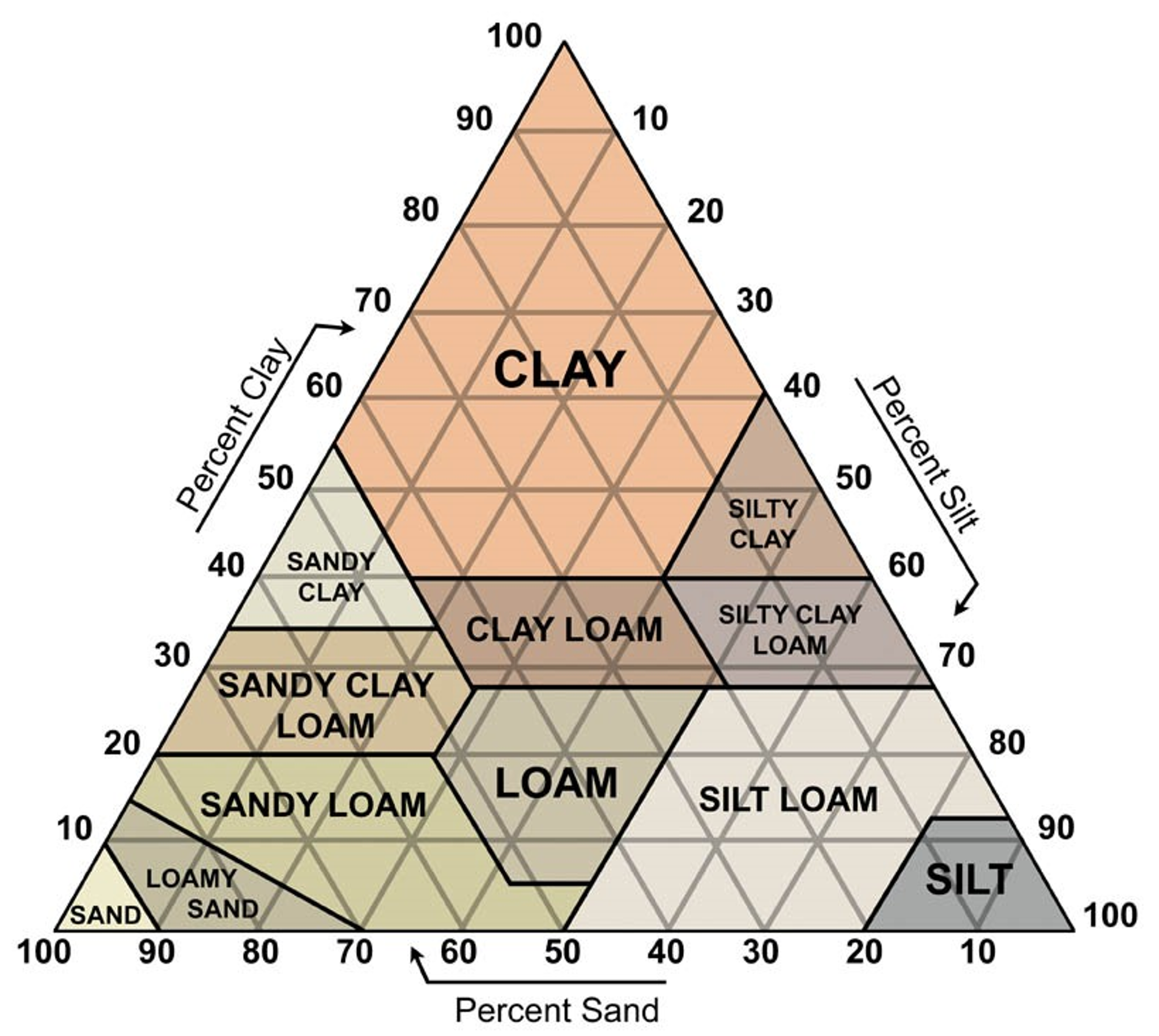
  • The soil composition did not vary as drastically as last samples. It did contain 4 different layers of the soil, but the fourth dark top layer was just additional organic matter that was not poured out.  It roughly had 1.5-mL of sand, 1.0-mL of silt, and 0.5-mL of clay. Making it 50% sand, 33.33% silt, and 16.67% clay.
  • Soil Composition 09/24/18 Bottom Layer: Sand Dark Middle Layer: Silt Light Layer: Clay

  • LB broth before use was not contaminated, no cloudiness was reported and there was no precipitate in the solution at the time of use.
  • The mass of the dry soil was 6.21 grams after being left out all weekend to encourage water evaporation. Therefore making the mass of water 3.91 grams.

Analysis/Conclusions:

  • Based off graphic below and the percentages of the soil composition obtained from the experiment, the best estimate for the soil type is Loam. Loam is a fertile soil with a roughly 40-20-20 mineral composition of sand-silt-clay, which is very close to the results gathered in the experiment.
  • Image result for sand silt clay
  • By using the water percentage equation, the final calculated percentage of water in the soil was 24.80%. This is only 4% more water than the previous soil sample which was gardeners soil. Possibly this increase in water will be beneficial for the presence of phage.

    Work for calculating water percentage


Next Steps:

  • Analyze the plaque assay results for the presence of phage. If there are plaques present, a serial dilution will be performed to get a high titer of phage. If no plaques are present, a second plaque assay will be performed.
September 27

Plaque Assay for Original Plaque (Redo)

Print Friendly, PDF & Email

Title: Plaque Assay for Original Plaque (Redo)

Date: 26 September 2018

Rationale: Due to an error with the Arthrobacter culture, the previous experiment’s results are rendered invalid. The bacteria used was not Arthrobacter, therefore an Arthrobacterphage couldn’t grow on a bacterial lawn. Therefore the experiment is repeated. The attached pictures show the previous (contaminated/invalid) plaque assays.

Procedure: Aseptic zone created by washing the lab bench with CiDecon and ethanol and a heat lamp was lit.

  • The picked 10^0 lysate from the previous experiment was used for 2 more 10^0 plaque assays.

The following recipe was used to make 8 plaque assays (7 PA + 1 Top Agar Control):

  • 18.9 mL LB Broth
  • 180 microliters CaCl2
  • 20 mL 2x TA

4.6 mL pipetted into a vial containing 0.4 mL new Arthrobacter strain + 10 microliters 10^0 lysate. The mixture was plated, cooled for 10-15 minutes, and incubated. \

Conclusions: Much like the previous experiment, both plaque assays must pass in order to continue passaging. If the test fails, soil will be recollected and the washing/purification process will start over.

September 27

09/26/18- Purification run 2 repeated

Print Friendly, PDF & Email

09/26/18

Objective:

  • Make another plaque assay from the extracted phages from the plate from the first serial dilution

Pre-Lab Observations:

  • After checking the positive control made by the lab instructors, it was discovered that the arthrobacter culture used on 09/24/18 was not actually arthrobacter.
  • So, new plaque assays must be made from the same phage extract used on 09/24/18
  • The control plate from the plaque assays prepared on 09/24/18 was not contaminated.

Procedure:

  1. Cidecon was poured on the desk and wiped till the desk was dry. Then, 70% ethanol was poured and wiped until it was all over the table and then it was allowed to evaporate. After the ethanol had evaporated, an ethanol lamp was lit, setting up the aseptic zone.
  2. 400 μl of arthrobacter was retrieved from the lab instructor
  3. Using the micropipette, 10 microliters of the 10^0 bacteriophage mixture was transferred to the arthrobacter vial.
  4. The vial was then allowed to rest on the test tube rack for 15 minutes
  5. While the vial was resting, one Top Agar mixture was made for the group.
  6. The LB broth was retrieved from its storage bath, along with a 50 ml conical tube and a serological pipette
  7. While in the aseptic zone, 8.4 ml of LB broth was transferred to the 50 ml conical vial.
  8. Then, 1 M CaCl2 stock solution was retrieved from the lab instructor.
  9.  Using the micropipette, 90 microliters of the CaCl2  was transferred to the 50 ml conical tube with the LB broth.
  10. The vial was then set on the rack.
  11. 10 ml of the 2X TA was added to the LB broth and Cacl2 after the sample was allowed to enrich the arthrobacter for 15 minutes.
  12. Using another serological pipette, 4.5 ml of the top agar mixture was transferred to the test tube with the arthrobacter and the phage extract.
  13. The contents of the test tube were then poured onto the agar plate.
  14.  Part of the top agar mixture was poured into the top agar control plate for the group.
  15. To let the top agar solidify, the plates were allowed to rest for 15 minutes.
  16. The plates were placed upside down in the incubator, where they will remain for 48 hours

Analysis and Conclusion:

The procedures were properly performed in the aseptic zone and the chances on contamination were minimized. the control plate from 09/24/18 was not contaminated, which was a surprising outcome due to the repeated contamination of the control plates in previous spot tests and plaque assays. it may have been the plates that were contaminated when contaminated control plates were a result because more caution was used while picking plates for assays on Monday.