September 28

9/24/18 Phage Purification Passage 2

9/24/18 Passage #2 of Enriched Lysate from Soil Sample #2

Objective:

The goal of this procedure is to passage our phage a second time as part of the phage purification process. Last lab we picked a potential plaque, preformed dilutions and the did multiple assays. This could be called the “first passage” (out of three). This lab we will pick the most promising plaque and do another assay (passage two). By passaging our phage we seek to isolate and purify 1 specific strand of phage. Once we do this (following the process found in the image below), we can move on to experimenting with our specific phage strain.

We are also seeking to avoid contamination even further, as can be seen in the analysis section of this report our previous TA control was contaminated.

We are also seeking to address the following questions every lab:

The overarching question this test seeks to address is: Is the presence of phage determined by species of oak tree from which soil was collected?

In other words, are specific oak tree species more likely to have Arthrobacter bacteria phages in the soil surrounding them?

The question specific to my lab table is: Is the a difference in the presence of phage between live oaks and red oaks on Baylor’s campus?

As a group we hope to expand our question to include more species as we gather data so that we can better address our overarching question and we will look at our metadata to examine weather or not there are other factors that may determine phage presence.

Procedures and Protocols:

Materials for Aseptic Zone:

  • CiDecon
  • 70% Ethanol
  • Ethanol Burner

Materials for Plaque Assay:

  • .5 ml Arthrobacter
  • incubator
  • Pipette
  • Test tube stand
  • 50 ml tubes
  • Culture tube
  • LB Broth
  • 2X TA
  • 1M Calcium Chloride
  • Agar plate
  • Serological pipette

Materials for Phage Picking:

  • Agar plates with plaques of interest
  • Micropipette tip
  • Phage buffer
  • Microcentrifuge tubes (incorrectly referred to as pipette caps in previous entries)

In order to complete the procedure an aseptic zone was created.

  1. CiDecon was applied to the lab table with a squeeze bottle and wiped away with a paper towel
  2. 70% Ethanol was also applied with a squeeze bottle, spread with a paper towel, and allow to evaporate
  3. An ethanol burner was light in order to use the rising heat from the flame to form the aseptic zone

Then a phage was picked *Note: Each group member picked the most promising plaque from their respective plates for a total of 3 picked plaques*

  1. 100 µL of phage buffer was transferred into a microcentrifuge tube labeled with initials, date and the description “P2” (P2 meaning passage 2)
  2. Four plaques were deemed to be promising, one was selected for a plaque assay, and the other three were selected for later spot test (image below) 
  3. A pipette tip was used to stab the center of the chosen plaque on each plate (the chosen plaque is indicated my the red arrow on the image below) *Note: My hands shake and it is possible I contaminated my pipette tip with the surrounding agar when I tried to stab my plaque; however, it does not appear that I contaminated my last picked plaque so I will assume I did not contaminate this one ether* 
  4. The phage-infected tip was swirled in the phage buffer and then the solution was vortexed and set aside.

Then a plaque assay on solution in the tube labeled “P2” was preformed

  1. Four agar plates were gathered and set aside.
  2. The agar was prepared according to the following recipe (makes four plates):
  3. Under aseptic conditions, 8.o ml of LB broth was transfered into a 50 ml tube.
  4. Under aseptic conditions, 90 µL of 1 M CaCl2 was transferred into the same 50 ml tube.
  5. 10 µL of the P2 lysate was aseptically transferred into a culture tube containing .5 ml of Arthrobacter using a Serological pipette
  6. The culture tube was capped and set aside for 15 minutes. This process was repeated twice more (once for each group member). *Note: this was a mistake from the usual process because we forgot to put the lysate into the culture tubes before we started prepping the agar, so after the first two steps of agar prep we had to stop and wait the 15 minute incubation period*

The lysate and bacteria were allowed to sit in the culture tube for 15 minutes

  1. Under aseptic conditions,  5.o ml of 2X TA was transferred into the same 50 ml tube
  2. The mixture was pipetted several times to mix it
  3. Three agar plates were labeled with initials, date, and “P2” while a forth was labeled with the date and description “TA control”
  4. 4.5 ml of the contents in the 50 ml tube was transferred to the plate labeled “TA control”
  5. The plate was swirled and set aside
  6. 4.5 ml of the contents in the 50 ml tube was transferred into the culture tube containing lysate and bacteria
  7. The mixture was pipetted several times to mix it
  8. This process was repeated twice more, once for each group memeber
  9. Then the mixture was poured from the culture tube into the agar plate labeled with initials, date, and description
  10. The plate was swirled and then set aside for 10 minutes to allow agar to solidify. This procedure was repeated twice more, once for each group member. *Note: when swirling Aman’s plate some of the liquid agar spilled out of his plate, potentially disrupting his results* 
  11. Once the labeled plaque assay had solidified, the plate was inverted and placed in the incubator
  12. Plates were left to incubate until nest class
Results:

The results of the plaque assay on the P2 lysate will be recorder here when available. It is reasonable to assume though, that there will be plaques as there have been plaques in the two previous assays.

Update:

There were no identifiable plaques on our plates on Wednesday (see plate below), but after our TA’s ran a control plate they discovered that during this lab we did not plate with artho. This makes all of our results from this lab invalid/unusable, and it explains why there were no plaques to be found. We will infer (and confirm with later testing) that there is still viable phage in our P2 tube, and as we continue to passage, it should be one strain and only one strain.


Analysis:

The results of this lab are invalid because we did not use arthro to plate our phage; however, there are still several valuable things that can be inferred from this. The first thing that this lab demonstrated is that there are a lot of ways for things to become contaminated. The image above shows the control plate from the lab conducted on 9/19/18. The plaques from that assay were picked to run the procedures detailed above, so it is important to understand forms of contamination. In this case we knew that we did not contaminate our sample and that our broth was not contaminated because we took extra precautions on 9/19. Based on how the contaminated appear to spread, we inferred that the contamination came from the plate itself, teaching us to more fully examine our agar plates before using them. In addition, based on the mistake with the arthro, we learned that mistakes can happen in every section of the scientific process and that we should always be careful.

Future:

Had our procedure gone to plan, we would have preformed passage #3 on Wednesday, but because we didn’t plate with arthro, we will redo our passage #2 on Wednesday, following roughly the same procedure, being careful to make the same mistakes.

September 21

9/17/18 Plaque Assay of Soil #2 Enriched Lysate and Collection of Soil Sample #3

9/17/18 Plaque Assay of Soil #2 Enriched Lysate and Collection of Soil Sample #3

Objective:

The goal of these procedures is to use the previously created lysate to preform a plaque assay to test for phage presence in our previously collected soil (soil sample #2). In addition, during this lab period we also collected a third soil sample in case the plaque assay on soil sample two yielded a negative result. Our goal is to isolate a phage, and because our spot test yielded negative results, we did a plaque assay to confirm while also preparing to take next steps if soil sample 2 is negative for phage. We are also trying to avoid the contamination that occurred on our controls (see below). We are also seeking to address the following questions every lab:

The overarching question this test seeks to address is: Is the presence of phage determined by species of oak tree from which soil was collected?

In other words, are specific oak tree species more likely to have Arthrobacter bacteria phages in the soil surrounding them?

The question specific to my lab table is: Is the a difference in the presence of phage between live oaks and red oaks on Baylor’s campus?

As a group we hope to expand our question to include more species as we gather data so that we can better address our overarching question and we will look at our metadata to examine weather or not there are other factors that may determine phage presence.

In addiction to our guiding questions, for this lab we were asked to consider the following:

  1. Group 4 all had plaques on their plaque assays. Justin had the most and well defined plaque (but all 3 got plaque). They each did a spot test in addition to their plaque assays, but only Justin had a plaque on his spot test. What do you think is going on?
    • After considering this question we determined that Justin’s sample likely had the highest titer and therefore resulted in more clearing and better defined spots. It is also possible that the way he cared for his sample was more conducive to phage survival (ether during soil transport or enrichment).
  2. Lathan checked a purified lysate by a plaque assay using 10 µL of a 10^-3 lysate. He got 14 plaques. How many µL of Lathan’s lysate should he add to web a plate (8 cm in diameter) if his average plaque diameter is 1 mm.
    • 4.6 µL of 10^0 lysate (work below)
Procedures and Protocols:

Materials for Aseptic Zone:

  • CiDecon
  • 70% Ethanol
  • Ethanol Burner

Materials for Plaque Assay:

  • .5 ml Arthrobacter
  • incubator
  • Pipette
  • Test tube stand
  • 50 ml tubes
  • Culture tube
  • LB Broth
  • 2X TA
  • 1M Calcium Chloride
  • Agar plate
  • Serological pipette

Materials for Soil Collection:

  • Sharpie
  • Tape Measure
  • Plastic Bags
  • Digging tools
  • 15 ml Tubes

In order to complete the procedure an aseptic zone was created.

  1. CiDecon was applied to the lab table with a squeeze bottle and wiped away with a paper towel
  2. 70% Ethanol was also applied with a squeeze bottle, spread with a paper towel, and allow to evaporate
  3. An ethanol burner was light in order to use the rising heat from the flame to form the aseptic zone

Then the plaque assay on the enriched lysate from soil sample #2 was preformed.

  1. Four agar plates were labeled. An agar plate was labeled with initials, date, and description for each group member, and one agar plate was labeled with data and “TA control”
  2. The remaining enriched lysate from last procedure (spot test), stored in a pipette cap, was gathered (like cap picture below)
  3. 10 µL of the remaining enriched lysate was aseptically transferred into a culture tube containing .5 ml of Arthrobacter using a Serological pipette
  4. The culture tube was capped and set aside for 15 minutes. This process was repeated twice more (once for each group member).

While the lysate and bacteria are allowed to sit in the culture tube the agar was prepared.

  1. The agar was prepared according to the following recipe (makes four plates):
  2. Under aseptic conditions, 8.o ml of LB broth was transfered into a 50 ml tube.
  3. Under aseptic conditions, 90 µL of 1 M CaCl2 was transferred into the same 50 ml tube. *Note: the first time we attempted to pipette the calcium, we accidentally drew too much out so we had to discard the pipette tip full of calcium and try again*
  4. Under aseptic conditions,  5.o ml of 2X TA was transferred into the same 50 ml tube
  5. The mixture was pipetted several times to mix it

When agar preparations were finished the bacteria and lysate had been allowed to sit for 15 minutes

  1. 4.5 ml of the contents in the 50 ml tube was transferred into the culture tube containing lysate and bacteria
  2. The mixture was pipetted several times to mix it
  3. Then the mixture was poured from the culture tube into the agar plate labeled with initials, date, and description
  4. The plate was capped and set aside for 10 minutes to allow agar to solidify. This procedure was repeated twice more, once for each group member.
  5. The remaining contents in the 50 ml tube was poured onto the TA control agar plate
  6. The TA control plate was allowed to sit for about 10 minutes before being placed into the incubator
  7. Once the labeled plaque assay had solidified, the plate was inverted and placed in the incubator
  8. Plates were left to incubate until nest class

Soil collection was preformed as follows:

  1. The species of tree that soil should be collected from was identified *Note: in this case we choose live oak as we did in soil sample 2*
  2. The provided bags were labeled with name, date, and description
  3. A tree of this species was loacted on campus
  4. The GPS coordinates were noted
  5. The tree’s trunk diameter was measured and recorder
  6. The tree’s height was measured and recorded using shadow length of a known height and then measuring the shadow of the tree and using trig
  7. The tree’s average canopy diameter was measured and recorded by taking measurement of the shortest and longest diameters and averaging them together (results in the table in results column)
  8. A digging instrument was used to clear debris and dig down several centimeters into the soil
  9. The provided plastic bags were filled with soil and a tree leaf (for confirmation of species identification)
  10. The bags were brought back to lab
  11. Refrigerate Tube and bag until next class
Results:

The results immediately gathered from these procedures are seen in the table above. The results of the plaque assay on soil #2 will be recorder here when available. It will not be possible to ascertain weather or not phage are present in soil sample #3 until the soil can be washed and then assays and spot tests can be preformed.

Update:

There are visible plaques on all three plates (see images below for my plates), but their is also contamination in the control. We are unable to determine how this contamination came to be, but we are operating under the assumption that it is not the cause of plaques forming. We will test this assumption in the future.

Analysis:

Based on the appearance of the tree and the measurements we took, we would assert that the tree is most likely healthy. Should we find phage in this soil and in the soil of trees nearby, this information could become important in determining if tree health is a factor for phage presence. It is also important to note that this was a live oak tree and it could become clear that live oak trees do not readily have phage in their soil after further sailing and analysis of soil sample #2’s plaque assay. The opposite could also be true and we will be more equipped to analyse our data once we test for phages.

Update:

Based on the results of visible plaques we can assert that there is likely phage present in the soil from sample #2. However, because the control plate was contaminated we cannot be certain. The results of further testing of plaques should reveal if there are in fact phages in soil sample #2, or if my group simply managed to contaminate everything

.

Future:

If necessary I will wash the that soil was collected (soil #3) and create lysates in order to run spot tests and plaque assays.

Based on the update, this will not be immediately necessary. On Wednesday I will pick a plaque and preform serial dilutions. Based on the results of that I will likely try to web a plate or be forced to begin testing soil sample #3.

September 14

9/10/18 Soil Washing and Metadata Collection

9/10/18 Soil Washing and Metadata Collection

Objective:

The goal of this procedure was to wash and enrich the new soil that was collected previously and to collect other soil metadata. We did so that we could address our questions and begin table level study of phages in soil.

The overarching question this test seeks to address is: Is the presence of phage determined by species of oak tree from which soil was collected?

In other words, are specific oak tree species more likely to have Arthrobacter bacteria phages in the soil surrounding them?

The question specific to my lab table is: Is the a difference in the presence of phage between live oaks and red oaks on Baylor’s campus?

As a group we hope to expand our question to include more species as we gather data so that we can better address our overarching question.

Procedures and Protocols:

Materials for Aseptic zone:

  • CiDecon
  • 70% Ethanol
  • Ethanol Burner

Materials For Soil Washing:

  • Syringe filter
  • .5 ml Arthrobacter
  • 50 ml conical vial
  • 15 ml conical vial
  • LB Broth
  • refrigerator
  • Incubator
  • Centrifuge
  • Pipette
  • Scale
  • Weigh Boat
  • Test tube stand

Materials for % Water Analysis:

  • Scale
  • Weigh Boat

Materials for % Sand, Silt, Clay Analysis:

  • Falcon Tube
  • Dispersion Fluid
  • Deionized (DI) water

Materials for pH Test:

  • pH vial
  • DI water
  • pH Paper
  • pH comparison color scale

In order to complete the procedure an aseptic zone was created:

  1. Clean off the work space (lab table) with CiDecon applied with a squeeze bottle and wiped away with a paper towel
  2. Apply 70% Ethanol with a squeeze bottle, spread with a paper towel, and allow to evaporate
  3. Light an ethanol burner in order to use the rising heat from the flame to form the aseptic zone

*The soil washing and enrichment was conducted while other tests were being conducted simultaneously by other group members; for the sake of a simple procedural explanation, all of the steps of each of the following procedure will be detailed separately. If there was an error or if the process of one procedure effected another it will be noted*

The soil was washed and enriched according to the following procedure:

  1. ~2 ml of soil was placed into a 15 ml conical vial
  2. 10 ml of LB broth was added, bring the combined contents of the vial to the 10.5 ml mark
  3. The vial was shaken and vortexed intermittently for 15 minutes
  4. The vial was then massed and centrifuged at 10,000 g
  5. After centrifugation a syringe filter was to separate the lysate from the remaining sample
  6. ~9 ml of lysate was filtered  with syringe filter into the 50 ml conical *Note this is less than the recommended 10 ml*
  7.  ~1 ml of lysate was filtered  with syringe filter into the 15 ml conical
  8. .5 ml of Arthrobacter was added to the 9 ml of lysate in the 50 ml conical and put in the incubator until next class ( 47 hours)
  9. The remaining 1 ml of lysate in the 15 ml conical was put in the fridge

% Water analysis was preformed according to the following procedure:

  1. A weigh boat was weighed and the mass was recorded (see table in results)
  2. Then a small amount of dirt was poured into the weigh boat and the combined weight was recorder (see table)
  3. The weigh boat was labeled with initials and date and allowed to sit in the fume hood until next lab (~48 hours)

% Sand, silt, clay analysis was preformed according to the following procedure:

  1. 10 ml of soil was put in a falcon tube using a tablespoon scoop
  2. DI water was added until the total contents of the the tube was 30 ml
  3. A piece of tape was attached to the tube and labeled with initials and date
  4. 3 drops of dispersion fluid were added to the tube
  5. The tube was covered with a hand and shaken for 30 seconds
  6. The tube was then placed under the fume hood until next lab (~48 hours)

The pH of the soil was collected according to the following procedure:

  1. A small amount of soil was scooped into a pH vial using a tablespoon scooper
  2. The rest of the vial was filled with DI wate
  3. The vial was shaken for 10 seconds
  4. Then the contents of the vial was allowed to settle for 2 minutes
  5. A strip of pH paper was put in the vial for 45 seconds and then compared to the pH color scale

(The image shows the resulting color change of the DI water after the pH slip was removed)

Results:

The majority of these procedures will not have results until next lab and this entry will be updated when results are available. That said the pH of the tested soil sample was between 6.0 and 6.5 leading us to estimate a pH of 6.2.

In addition the soil washing seems to have gone well and has resulted in both enriched and direct lysate for future testing.

Update:

Analysis:

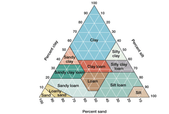
These procedure were meant to learn more about our new soil samples and get us prepared for future testing. Based on our results I can assert that my soil sample is very sandy and using the chart seen below I can assert that the soil is sand or potentially sandy loam if enough material was still in suspension. I was also assert that my soil sample was slightly acidic based on the results of my pH test. This will help my group address our question further because having a collection of soil metadata will help us determine weather or not other factors besides tree species determine phage presence.

Image result for soil pyramid(https://samanthaapes.weebly.com/apes-in-a-box-soil-pyramid.html)

Future:

This entry has been updated to reflect the results of the metadata testing; however, when the procedures stated above were completed the initial future procedures were to simply check on the results of the experiments after the appropriate amount of time had elapsed. Now that this is completed spot tests will be preformed using the lysate created using the above procedures and depending on the results of those spot tests plaque assays or more soil collection will be preformed.

September 7

9/5/18 Soil Sample Collection Live Oak

9/5/18 Soil Sample Collection Live Oak

Objective:

The goal of this procedure is to collect new soil to wash and test for the presence of phage. The last soil sample that was collected appear to be negative for phage after two failed assays, necessitating new soil collection. In addition, new soil collection was also necessary in order to answer our overarching and table specific questions.

The overarching question this test seeks to address is: Is the presence of phage determined by species of oak tree from which soil was collected?

In other words, are specific oak tree species more likely to have Arthrobacter bacteria phages in the soil surrounding them?

The question specific to my lab table is: Is the a difference in the presence of phage between live oaks and red oaks on Baylor’s campus?

As a group we hope to expand our question to include more species as we gather data so that we can better address our overarching question.

Procedures and Protocols:

Materials:

  • Sharpie
  • Tape Measure
  • Plastic Bags
  • Digging tools
  • 15 ml Tubes

Soil collection is preformed as follows:

  1. Identify the species of tree soil should be collected from
  2. Locate a tree of this species on campus *Note: We settled on live oak*
  3. Make note of the GPS coordinates 
  4. Measure and record the tree’s trunk diameter
  5. Measure and record the tree’s height be using shadow length of a known height and then measuring the shadow of the tree and using trig
  6. Measure and record the tree’s average canopy diameter by taking measurement of the shortest and longest diameters and average them together (results in the table in results column)
  7. Using digging instrument to clear debris and dig down several centimeters into the soil
  8. Fill the provided plastic bag with soil and a tree leaf (for confirmation of species identification)
  9. Label bag with name, date, and description
  10. Bring bag back to lab and put ~2 ml of soil into the 15 ml tube
  11. Refrigerate Tube and bag until next class
Results:

The results immediately gathered from this procedure are seen in the table above. It will not be possible to ascertain weather or not phage are present in the soil until the soil can be washed and then assays and spot tests can be preformed.

Analysis:

There is very little data that can be analyzed. However, based on the appearance of the tree and the measurements we took, we would assert that the tree is most likely healthy. Should we find phage in this soil and in the soil of trees nearby, this information could become important if tree health is a factor for phage presence. It is also important to note that this was a live oak tree and it could become clear that live oak trees do not readily have phage in their soil after further sailing. The opposite could also be true and we will be more equipped to analyse our data once we test for phages.

Future:

On Monday’s lab I will wash the that soil collected, and in the following lab periods I will likely run spot tests and plaque assays on the lysate from the soil.

September 7

8/31/18 Plaque Assay of Enriched Lysate Attempt Two

8/31/18 Plaque Assay of Enriched Lysate Attempt Two

Objective:

The goal of this procedure is to determine weather or not bacteria phages are present in the soil collected last week. Based on the results of the spot test it is possible that there are phages, but the previous plaque test was contaminated (see below) so now this assay will be conducted to recheck.

The overarching question this test seeks to address is: Is the presence of phage determined by species of oak tree from which soil was collected?

In other words, are specific oak tree species more likely to have Arthrobacter bacteria phages in the soil surrounding them?

The question specific to my lab table is: Is the a difference in the presence of phage between live oaks and red oaks on Baylor’s campus?

As a group we hope to expand our question to include more species as we gather data so that we can better address our overarching question.

Procedures and Protocols:

Materials:

  • CiDecon
  • 70% Ethanol
  • Ethanol Burner
  • .5 ml Arthrobacter
  • incubator
  • Pipette
  • Test tube stand
  • 50 ml tubes
  • 15 ml tube
  • LB Broth
  • 2X TA
  • 1M Calcium Chloride
  • Agar plate
  • Serological pipette

In order to complete the procedure an aseptic zone was created.

  1. Clean off the work space (lab table) with CiDecon applied with a squeeze bottle and wiped away with a paper towel
  2. Apply 70% Ethanol with a squeeze bottle, spread with a paper towel, and allow to evaporate
  3. Light an ethanol burner in order to use the rising heat from the flame to form the aseptic zone

Then the plaque assay on the enriched lysate was preformed.

  1. Label two agar plates. Label one with initials, date, and description “TA control”
  2. Label a second agar plate with initials, date, and description “Enriched Lysate Assay (HB)”
  3. Gather the remaining enriched lysate from last procedure (found in the pipette cap seen below)
  4. Using a Serological pipette, aseptically transfer 10 µL of the remaining enriched lysate into a 15 ml tube containing .5 ml of Arthrobacter
  5. Reseal tube and recap pipette cap
  6. Allow the lysate and bacteria solution to sit for 15 minutes while the agar is prepared

While the lysate and bacteria are allowed to sit in the culture tube prepare the agar

  1. Prepare the agar according to the following recipe (makes two plates):
  2. Under aseptic conditions, pipette 5.o ml of LB broth into a 50 ml tube. Cap the tube. *Note: This was a mistake that will be addressed later in the procedure*
  3. Under aseptic conditions, pipette 45 µL of 1 M CaCl2 into the same 50 ml tube. Cap the tube.
  4. Discard 50 ml tube with LB broth and CaCl2 because the measurements were wrong
  5. Under aseptic conditions, pipette 4.o ml of LB broth into a 50 ml tube. Cap the tube.
  6. Under aseptic conditions, pipette 45 µL of 1 M CaCl2 into the same 50 ml tube. Cap the tube.
  7. Under aseptic conditions, pipette 5.o ml of 2X TA into the same 50 ml tube
  8. Pipette the mixture several times to mix it

When agar preparations are finished the bacteria and lysate have been allowed to sit for 15 minutes

  1. Pipette 4.5 ml of the contents in the 50 ml tube into the lysate and bacteria 15 ml tube
  2. Pipette the mixture several times to mix it
  3. Pour the mixture from the 15 ml tube into the agar plate labeled with initials, date, and description “Enriched Lysate Assay (HB)”
  4. Cap the plate and allow the plaque assay agar to solidify for 10 minutes
  5. Aseptically Pipette 5 ml of the contents in the 50 ml tube onto the control TA plate
  6. Allow the TA control plate to sit for about 10 minutes before being placed into the incubator
  7. Once the labeled plaque assay has solidified, invert the plate and place it in the incubator
  8. Leave to incubate until next class (~94 hours)
Results:

The results of this procedure will not be immediately clear until Wednesday’s normal lab, but once they are available they will be included here.

Update: The top agar appears to have not solidified all the way because there is liquid in the plate. There do not appear to be any plaques in the areas where that agar remained intact suggesting a negative assay.

Analysis:

Because the Agar did not stay solidified it is hard to analyse these results with certainty; however, because there did not appear to be plaques on the parts of the agar that remained solid it is likely that the assay was negative. This suggests that there were not phages in the soil collected, or that there are not enough to cause results.

Future:

Due to the two unsuccessful assays, I will be collecting soil next lab in an attempt to find viable phage for isolation and purification.

August 31

8/29/18 Plaque Assay of Enriched Lysate

8/29/18 Plaque Assay of Enriched Lysate

Objective:

The goal of this procedure is to determine weather or not bacteria phages are present in the soil collected last week. Based on the results of the spot test it is likely that there are phages present (see spot test results below). In this procedure a plaque assay on the enriched lysate will be conducted to confirm the results of the spot test.

The overarching question this test seeks to address is: Is the presence of phage determined by species of oak tree from which soil was collected?

In other words, are specific oak tree species more likely to have Arthrobacter bacteria phages in the soil surrounding them?

Procedures and Protocols:

Materials:

  • CiDecon
  • 70% Ethanol
  • Ethanol Burner
  • .5 ml Arthrobacter
  • refrigerator
  • Pipette
  • Test tube stand
  • 50 ml tubes
  • LB Broth
  • 2X TA
  • 1M Calcium Chloride
  • Agar plate
  • Serological pipette

In order to complete the procedure an aseptic zone was created.

  1. Clean off the work space (lab table) with CiDecon applied with a squeeze bottle and wiped away with a paper towel
  2. Apply 70% Ethanol with a squeeze bottle, spread with a paper towel, and allow to evaporate
  3. Light an ethanol burner in order to use the rising heat from the flame to form the aseptic zone

Then the plaque assay on the enriched lysate was preformed.

  1. Divide the bottom of an Agar plate into four sections and label them Groups 1, 2, 5, and 6 in order to create a TA controlSet aside.
  2. Label a second agar plate with initials, date, and description (plaque assay will be preformed in this plate).
  3. Gather the remaining enriched lysate from last procedure (found in the pipette cap seen below)
  4. Using a Serological pipette, aseptically transfer 10 µL of the remaining enriched lysate into a culture tube containing .5 ml of Arthrobacter
  5. Reseal tube and recap pipette cap
  6. Allow the lysate and bacteria solution to sit for 15 minutes while the agar is prepared

While the lysate and bacteria are allowed to sit in the culture tube prepare the agar *Note:this was a collaborative effort so the agar was prepared for three full agar plates and a control:

  1. Prepare the agar according to the following recipe (use 4 plate recipe):
  2. Under aseptic conditions, pipette 8.o ml of LB broth into a 50 ml tube. Cap the tube.
  3. Under aseptic conditions, pipette 90 µL of 1 M CaCl2 into the same 50 ml tube. Cap the tube.
  4. Under aseptic conditions, pipette 10.o ml of 2X TA into the same 50 ml tube
  5. Pipette the mixture several times to mix it

When agar preparations are finished the bacteria and lysate have been allowed to sit for 15 minutes

  1. Pipette 4.5 ml of the contents in the 50 ml tube into the lysate and bacteria culture tube
  2. Pipette the mixture several times to mix it *Note: in the process of pipetting the mixture and air bubble caused some of the mixture to spill out of the culture tube, potentially upsetting the ratios in place*
  3. Pour the mixture in the culture tube into the agar plate labeled with initials, date, and description
  4. Cap the plate and allow the plaque assay agar to solidify for 10 minutes
  5. Aseptically Pipette 1 ml of the contents in the 50 ml tube onto the control TA plate in the section labeled group 6
  6. Allow the TA control plate to sit for about 10 minutes before being placed into the incubator
  7. Once the labeled plaque assay has solidified, invert the plate and place it in the incubator
  8. Leave to incubate until next class
Results:

The results of this procedure will not be immediately clear until Friday’s open lab or Wednesday’s normal lab, but once they are available they will be included here.

Update: The plates appear to be inconclusive based on cloudy appearance and due to a lack of positive or negative controls, one cannot make any assertions about phage presence.

Analysis:

It is hard to analyse the results of this lab because the results themselves are unclear. However, it can be concluded that when the agar plates are redone thicker agar lawns should be used and i would be beneficial to include positive and negative controls as a means of comparison. It is also possible that samples were contaminated, but it is hard to confirm this.

Future:

Due to the inconclusive nature of the plaque assays, I will need to redo my plaque assay likely with positive and negative controls in order to confirm the results of my spot test.

August 31

8/27/18 Spot Test

8/27/18 Spot Test

Objective:

The goal of this procedure is to determine weather or not bacteria phages were present in the soil collected last week. This will be determined by using small amounts of lysate on auger plates to look for plaques that would indicate phage particles. By performing this test

Procedures and Protocols:

Materials:

  • CiDecon
  • 70% Ethanol
  • Ethanol Burner
  • .5 ml Arthrobacter
  • refrigerator
  • Pipette
  • Test tube stand
  • 50 ml tubes
  • LB Broth
  • 2X TA
  • 1M Calcium Chloride
  • Agar plate
  • Micropipette

In order to complete the procedure an aseptic zone was created.

  1. Clean off the work space (lab table) with CiDecon applied with a squeeze bottle and wiped away with a paper towel
  2. Apply 70% Ethanol with a squeeze bottle, spread with a paper towel, and allow to evaporate
  3. Light an ethanol burner in order to use the rising heat from the flame to form the aseptic zone

Then the spot test could be preformed.

  1. Divide the bottom of an Agar plate into three sections and label them E for enriched isolation, D for direct isolation, and B for phage buffer *note that this was a collaborative effort to some degree so three agar plates were created*
  2. Create a separate Agar plate for a top agar (TA) control, label and set aside
  3. Gather previously created enriched isolation and direct isolation lysate
  4. Use a syringe to aseptically draw 3 ml of the enriched lysate out of the 50 ml tube
  5. Reseal tube
  6. Attach a filter to end of syringe and gently push 1.5 ml of lysate through the filter and into a pipette tip
  7. Cap the tip and set aside
  8. Set aside two 50 ml tubes and label them as TA control and Arthrobacter
  9. In the Arthrobacter tube add 13.ml of LB broth *The Arthrobacter tube was originally overfilled so some of the LB broth was pipette into the TA control tube*
  10. In the TA control tube add 4.5 ml of LB broth *Note that while adding LB broth to the TA tube the broth was spilled and the tube contents had to be emptied and new broth added*
  11. Set aside
  12. Add 42.75 μl Calcium Chloride and 5.0 ml of 2X TA to the TA control tube
    1. *The amount of calcium chloride to be added was determined based on the following calculations:
  13. Mix the contents of the TA control by shaking the tube vigorously *note that a pipette should have been used*
  14. Pipette 10 ml of the contents in the TA control tube into the agar plate labeled TA control
  15. cap plate and set aside for agar to solidify
  16. In the tube labeled Arthrobacter add 135 μl Calcium Chloride, 1.5 ml Arthrobacter, and 15.0 ml of 2X TA
  17. Use a pipette to mix solution by pulling it into the pipette and then expelling it several times
  18. Pipette 10 ml of solution from the Arthrobacter tube into each of the Agar plates
  19. On the Agar plate pour the remaining contents of the tube onto the plate because slightly less than 10 ml are left
  20. Allow Agar to solidify
  21. Using a Micropipette pipette 10 μl of filtered enriched lysate onto the section of plate labeled E, then pipette 10 μl of direct isolation lysate onto the section of plate labeled D, finally pipette 10 μl of phage buffer onto the section of plate labeled B
  22. Allow the plate to sit for about 10 minutes before being placed into the incubator
  23. Leave to incubate until next class (approximately 46 hours)
Results:

The results of this procedure will not be immediately clear until Wednesday’s lab, but once they are available they will be included here. Based on visible observations from lab today, the results of this procedure were four agar plates that appear to have been created correctly.

Update: There are visible plaques!!

Analysis:

Based on the Wednesday update, it can be inferred that there are bacteria phage present in my enriched and direct lysates. I will confirm this will a plaque update.

Future:

My future actions will heavily depend on the results of these spot tests. Regardless of weather or not plaques form though I will be doing plaque assay. So my next lab will detail that.

August 27

8/24/18 Enrichment One Part Two

8/24/18 Enrichment One Part Two

Objective:

The goal of this procedure was to finish preforming soil washing and enrichment on soil samples collected from oak trees around Baylor University’s campus in order to isolate bacteria-phages. The particular sample used in this procedure was found at 31*32’40” N 97*7’9″ W near Waco Hall. This is a continuation of 8/22/18 lab journal.

Procedures and Protocols:

Materials:

  • CiDecon
  • 70% Ethanol
  • Ethanol Burner
  • Top filter with 50 ml tube attached
  • Fume hood with a vacuum tube
  • .5 ml Arthrobacter
  • 15 ml conical vial
  • refrigerator
  • Pipette
  • Test tube stand
  • 50 ml tube

In order to complete the procedure an aseptic zone was created.

  1. Clean off the work space (lab table) with CiDecon applied with a squeeze bottle and wiped away with a paper towel
  2. Apply 70% Ethanol with a squeeze bottle, spread with a paper towel, and allow to evaporate
  3. Light an ethanol burner in order to use the rising heat from the flame to form the aseptic zone

Then the soil washing and enrichment procedure was complete (see photo below for full procedure and see last entry for previous steps).

  1. Remove the refrigerated sample (in a 50 ml tube) from the fridege
  2. Open the sealed top filter under the fume hood and attach to the vacuum tube
  3. Turn the vacuum on
  4. Use a small pipette to transfer the supernatant from the 50 ml tube to the top filter
  5. Wait for the supernatant to filter to a minimum of 10-15 ml (completely is preferred and 18 ml were collected in this instance)
  6. Once the desired about of lysate has been obtained discard the filter in the bio-hazard container and seal the 50 ml tube with the filtered lysate
  7. Under aseptic conditions pipette 5 ml of lysate into a 15 ml conical vile, label, and set aside
  8. Add .5 ml of Arthrobacter to the remaining lysate in the 50 ml tube *note that this tube should have been labeled but I forgot to do so* and seal the tube
  9. Place tube into shaking device and let it set until Monday

Important to note:

During this lab I was responsible for completing two separate soil washings, my own and my lab partners. I have only detailed my soil washing in the procedure above.

Results:

This procedure yielded a direct isolation (5 ml) and an enriched isolation (13 ml) of the supernatant. Both appeared of these isolations were yellow in color and could contain phages.

Analysis:

The procedure was more difficult than I initially assumed, and filter times were longer than expected. One possible way to improve upon the design of this lab could potentially be to centrifuge the soil and LB broth for a little bit longer in order to have better separation. It is possible, although unlikely that the results could have been contaminated during the course of this procedure and that may affect results. Assuming that this is not the case then future procedures will reveal weather or not phages are present.

Future:

My plans for the future are to use these two isolations to preform a spot test during Monday’s lab period. The results of this spot test will allow me to discover if there are phages present in the soil sample I collected.

August 27

8/22/18 Enrichment One Part One

8/22/18 Enrichment One Part One

Objective:

The goal of this procedure was to preform soil washing and enrichment on soil samples collected from oak trees around Baylor University’s campus in order to isolate bacteria-phages. The particular sample used in this procedure was found at 31*32’40” N 97*7’9″ W near Waco Hall.

Procedures and Protocols:

Materials:

  • CiDecon
  • 70% Ethanol
  • Ethanol Burner
  • 1 50 ml tube
  • ~ 20 ml LB Broth
  • Centrifuge

In order to accomplish this goal two procedures had to occur. The first was the creation of an aseptic zone.

  1. Clean off the work space (lab table) with CiDecon applied with a squeeze bottle and wiped away with a paper towel
  2. Apply 70% Ethanol with a squeeze bottle, spread with a paper towel, and allow to evaporate
  3. Light an ethanol burner in order to use the rising heat from the flame to form the aseptic zone

The second procedure was soil washing and enrichment (see photo below).

  1. Collect soil from designated oak tree by digging a few centimeters below the grass
  2. Place soil in a 50 ml tube up to the 15 ml line
  3. Add LB broth up to the 35 ml line under aseptic conditions
  4. Shake the tube for 15 minutes (during this 15 minutes the tube was dropped on the floor, potentially disturbing the soil and broth, but then the shaking resumed)
  5. Place tube in centrifuge for 5 minutes on 3,000 X g in order to separate the soil (pellet) and supernatant
  6. Place tube in the fridge to complete the procedure on a later date

Results:

This procedure yielded a tube of centrifuge separated supernatant and pellet as seen in the image. The solids separated by mass in the centrifuge with the most dense on the bottom. The centrifuge was spun on a low enough power to leave any existing phages in the supernatant.

Analysis:

The procedure had to be stopped halfway through so the results were incomplete and will be detailed in the next entry. Based on the results I do have I can make the assumption that the centrifuge didn’t fully separate my sample because it appears to be murky and there is debris on the top of the supernatant.

Future:

My plans for the future are to complete the procedure I started during Friday’s open lab in order to have an enriched sample and direct sample.