September 14

Washing and Determining the pH and Soil Components % (9/10/18)

Rationale: To perform a washing to separate the phages and bacteria in which the lysate will be divided into what will become enriched and direct isolation. To also determine the pH, % sand, % silt, and % clay

Materials:

  • 15 mL conical vial
  • 10 mL of LB broth
  • 20 mL of DI water
  • Soil Dispersion Liquid
  • Petri Dish
  • Syringe and Filters
  • Microcentrifuge tubes

Procedure:

  • Add 2 mL of Soil Sample 2 to a 15 mL conical vial.
  • Add LB broth up to the 12 mL mark in the vial.
  • Vortex the vial for five minutes, shake the vial for another five, and vortex the vial for the last five minutes.
  • While vial is on the vortex, add 10 mL of soil into a Falcon tube.
  • Add DI water up to the 30 mL mark in the Falcon tube.
  • Add three drops of soil dispersion liquid.
  • Shake the tube for 30 seconds.
  • Let the tub sit for 30 seconds.
  • The 15 mL vial now goes into the centrifuge at 10,000 g for 5 minutes.
  • While the 15 mL vial is in the centrifuge, weigh a petri dish then add soil and weigh it.
  • Set aside the petri dish with soil under a vacuum hood for 48 hours
  • FIlter the Soil Sample 2 supernatant using a syringe.
  • Use as many filters as needed to filter the supernatant.
  • Pour the lysate into a microcentrifuge tube labeled “Filtered Enriched 2.”
  • Take some soil from Soil Sample 2 and put it in a small glass vial to measure the pH.
  • Add some DI water and shake.
  • After shaking, take an inch of pH paper and put it in the mixture for about 45 seconds.
  • Take out the paper and determine the pH based on the color of the water at the top of the vial.
  • Dispose of the contents of the vial and clean it thoroughly.


Results and Analysis:

  • The weight of the Petri Dish: 7.19 g
  • The weight of the Petri Dish and soil: 11.80 g
  • pH of soil: 7.6
  • I had a lot of difficulty in filtering my supernatant using the syringe resulting in a shortage of lysate.


Soil Sample 2 immediately after shaking the Falcon Tube


Supernatant before filtering


Determination of pH by the color of the water


Conclusion and Future Plans:

  • To determine pH, remove a small sample of the soil sample and place it into a vial. Fill the vial up with water all the way to the rim. Shake the vial until the two components are evenly mixed. Take a piece of pH paper and determine the pH of the soil by using the color chart that is provided. Another procedure was to take some soil from Soil Sample 2 bag and to place it on a petri dish under a vacuum hood. The purpose is to determine the % water after 48 hours. The last procedure that was conducted is the filtering of the supernatant. Using a syringe and many filters, filtering the supernatant was a bit difficult and long due to the debris that was restricting the flow of the phages through the filter. Remove all the supernatant from the conical vial and filter for the phages which would go into a microcentrifuge tube labeled “Filtered Enriched 2.”
  • In the future, I plan to conduct a spot test to check for phages.
September 14

09/12/18 Plaque Assay, Spot Test, and Soil Metadata

Rationale:

The goal of today’s lab was to perform both a plaque assay and spot test as well as examine the soil metadata from the lab prior.

Materials:

  • LB Broth
  • 2X Top Agar
  • 2x 0.5 mL of Arthobacter
  • Calcium Chloride
  • Serological pipettes
  • Micropipettes
  • Balance

Procedure:

  1. This procedure began with establishing an aseptic zone using CiDecon to wipe the table down, 70% Ethanol to spray on the table and let evaporate, and a burner to create a convection current for air flow.
  2. I began with a spot test first, grabbing my enriched isolation labeled “GJA Enriched 2 09/10/18” and sent it off to centrifuge for 5 minutes at 3,000 g.
  3. While that was occurring, I found my soil sample that was left to evaporate and weighed it to find the mass of the dry soil and calculate the percent of water in the soil. This is the data below along with the equation used:
    Empty Petri Dish (mi) 6.90 g
    Petri Dish and Wet Soil (mf) 11.60 g
    Mass of Wet Soil (mwet soil) 4.70 g
    Petri Dish and Dry Soil (mf2) 10.62 g
    Mass of Dry Soil (mdry soil) 3.72 g
    Mass of Water (g) 0.98 g
    %Water 20.85%


  4. After calculating my percent water, I retrieved my enriched sample from the centrifuge and began to make the top agar by first adding 2.00-mL of LB Broth to a 50-mL conical tube labeled “GJA Top Agar 2 09/12/18”.
  5. Next I used a 20-200 µL micropipette to add 22.50-µL of Calcium Chloride to my LB Broth as well.
  6. Following this, I filtered approximately 1.5-mL of my enrichment through a 2 micron syringe filter and into a micro centrifuge tube and left that to sit while I transferred 0.5-mL of our Arthrobacter into my top agar solution.
  7. Once the bacteria was added, I used a serological pipette to add 2.5 mL of the 1x Top Agar solution. Then I swirled my 50-mL conical vial gently to allow everything to mix and I quickly poured the top Agar onto my plate and left it to solidify for 15 minutes.
  8. While my solution was solidifying, I examined my soil separation and poured off the supernatant so I could examine the sand, silt, clay composition of my soil.  Out of the 8-mL of soil present, 3.8-mL was sand, 2.5-ml was silt, and the remaining 1.7-mL was clay. This makes a percentage of 47.5% sand, 31.25% silt, and 21.25% clay
  9. After calculating the soil composition, my group decided to perform a plaque assay as well with the remaining time we had available.
  10. The group decided to make enough top agar for 3 plates plus a control. This meant we had 8-mL of LB Broth, 10-mL 2X TA, and 90-µL of Calcium Chloride.
  11. We began with adding the 8-mL of LB Broth with a serological pipette into one 50-mL conical vial.
  12. After adding the LB broth, the 90-µL of calcium chloride was added using a 10-100 µL micropipette.
  13. Once the calcium chloride was added, I combined 0.5-mL of Arthobacter with 10-µL of my lysate and let that infect for approximately 15 minutes.
  14. At the end of the 15 minutes, we added 10-mL of 2X Top Agar to the 50-mL conical vial through serological pipette. Immediately following this, I extracted 5-mL of the top agar solution, added it to a 15-mL conical vial with my arthrobacter and lysate solution, mixed it very quickly, and poured it into my plate to let it solidify for 15 minutes.
  15. With the remaining 5-mL of top agar left, we plated into our control plate
  16. Once the 15 minutes were up, the plates were immediately put into the incubator for 48 hours.

Data/Results

  • Water percentage of the soil was approximately 20.85%. This was moderately surprising because the soil when it was extracted was very wet, almost clay like.
  • The top agar came out looking very similar to previous top agars, despite my lysate being not as clear as everyone else’s.
  • The soil contains an extremely large amount of sand in comparison to silt and clay. This was unsurprising as our soil once dry was very crumbly and exhibited sand-like qualities.

Conclusions:

  • Soil was roughly 1/5 water. This could be a factor of its location to a nearby water source. There was also a man-made fountain nearby, which may contribute to the amount of water in the soil.
  • In addition to the water amount, the soil was a majority of sand present in the soil. This is most likely due to the dry location that the soil and tree is in.

Next Steps:

  • The next steps would be to analyze the plaque assay and spot test for the presence of phage in the soil. If there is phage present in the soil, then we will begin to analyze the amount of phage present. If there is no phage present, then I have to redo another plaque assay and spot test or gather a new sample.


September 14

9/12/18 Spot Test #2 and Completion of Metadata Collection

9/12/18 Spot Test #2 and Completion of Metadata Collection

Objective:

The goal of this procedure is to use the previously created lysate to preform a spot test to test for phage presence in our new collected soil. During this lab period the results of the metadata tests previously run will be collected in order to help us better answer our questions.

The overarching question this test seeks to address is: Is the presence of phage determined by species of oak tree from which soil was collected?

In other words, are specific oak tree species more likely to have Arthrobacter bacteria phages in the soil surrounding them?

The question specific to my lab table is: Is the a difference in the presence of phage between live oaks and red oaks on Baylor’s campus?

As a group we hope to expand our question to include more species as we gather data so that we can better address our overarching question and we will look at our metadata to examine weather or not there are other factors that may determine phage presence.

Procedures and Protocols:

Materials for Aseptic zone:

  • CiDecon
  • 70% Ethanol
  • Ethanol Burner

Materials For Spot test

  • .5 ml Arthrobacter
  • refrigerator
  • Pipette
  • Test tube stand
  • 50 ml tubes
  • LB Broth
  • 2X TA
  • 1M Calcium Chloride
  • Pipette cap
  • Phage Buffer
  • Agar plate
  • Micropipette
  • Syringe Filter

*In this lab period the results of soil metadata tests preformed last lab period were recorded. There were no procedural steps aside from recording information so the procedures for the metadata experiments will not be repeated here. However, the results of the metadata tests will be found both in this entry and the previous one to reflect both when they were collected and where they are applicable*

In order to complete the procedure an aseptic zone was created.

  1. Clean off the work space (lab table) with CiDecon applied with a squeeze bottle and wiped away with a paper towel
  2. Apply 70% Ethanol with a squeeze bottle, spread with a paper towel, and allow to evaporate
  3. Light an ethanol burner in order to use the rising heat from the flame to form the aseptic zone

Then the spot test could be preformed.

  1. Divide the bottom of an Agar plate into three sections and label enriched, direct, and PB for phage buffer *Note that the agar was made for four 5 ml plates*
  2. Create a separate Agar plate for a top agar (TA) control, label and set aside
  3. Gather previously created enriched lysate from incubator
  4. Mass lysate and centrifuge at 5,000 g for 5 minutes to pellet Arthrobactor
  5. Use a syringe to aseptically draw ~1.5 ml of the enriched lysate out of the 50 ml tube
  6. Reseal tube
  7. Attach a filter to end of syringe and gently push lysate through the filter and into a pipette tip
  8. Cap the tip and set aside
  9. Set aside a 50 ml tube
  10. In the 50 ml tube add 8.0 ml of LB broth
  11. Then add 90.0 μl Calcium Chloride
  12. Add 10.0 ml of 2X Top Agar and pipette to mix
  13. Add 4.5 ml of the mixture in the 50 ml tube to a culture tube and pipette to mix (repeat this twice more to create 3 culture tubes)
  14. Pour the contents of culture tube into labeled agar plate, swirl contents, cap plate and set aside for 10 minutes
  15. Pour the remaining liquid in the 50 ml tube into the agar plate labeled TA control
  16. Swirl contents, cap plate and set aside for agar to solidify (10 minutes) *Note: one of the members in my group found a possible containment in his agar plate so as a group we made a new control and new agar plate for him detailed below*
    1. Divide the bottom of an Agar plate into three sections and label enriched, direct, and PB for phage buffer *Note that the agar was made for two 5 ml plates*
    2. Create a separate Agar plate for a top agar (TA) control, label and set aside
    3. Set aside a 50 ml tube
    4. In the 50 ml tube add 4.0 ml of LB broth
    5. Then add 45.0 μl Calcium Chloride
    6. Add 5.0 ml of 2X Top Agar and pipette to mix
    7. Add 4.5 ml of the mixture in the 50 ml tube to a culture tube and pipette to mix
    8. Pour the contents of culture tube into labeled agar plate, swirl contents, cap plate and set aside for 10 minutes
    9. Pour the remaining liquid in the 50 ml tube into the agar plate labeled TA control
    10. Swirl contents, cap plate and set aside for agar to solidify
  17. Once agar has solidified use a Micropipette to pipette 10 μl of filtered enriched lysate onto the section of plate labeled enriched, then pipette 10 μl of direct isolation lysate onto the section of plate labeled direct, finally pipette 10 μl of phage buffer onto the section of plate labeled PB

  18. Allow the plate to sit for about 15 minutes before being placed into the incubator
  19. Leave to incubate until next class (approximately 120 hours)
Results:

The results of the spot test will not be discovered until Monday lab period and will be recorded in that lab entry. However, the results of the metadata experiments can be found below as they were collected during this lab period. The results show sand, acidic soil with a low water content.


Analysis:

The procedures conducted last lab and finished this lab are meant to provide more data that may inform future testing. Based on our results I can assert that my soil sample is sandy or potentially sandy loam if enough material was still in suspension. There is also a low percentage of water in the soil which could potentially affect if phage will be found. The information gathered  will help my group address our question further because having a collection of soil metadata will help us determine weather or not other factors besides tree species determine phage presence.

In addition, the results of the spot test will shed light on whether or not there is phage present in the soil we collected, which could help us address our question.

Future:

The next steps to be taken will depend on the results of the spot test. If the spot test is positive I will preform a plaque assay to confirm and the move on to phage isolation. If the test is negative, I will still preform a plaque assay to confirm, but I will likely also go collect new soil and begin the washing process again.

September 14

9/10/18 Washing of Soil B and Metadata

Objective:

  • Wash the soil sample (Soil B) to remove unnecessary material, such as dirt, rocks, and bacteria.
  • Isolate possible bacteriophage found in the soil, in lysate.
  • Collect metadata on soil sample “Soil B” to obtain more information about the environment the soil was collected in and have further data on the preferred environment of Arthrobacter phage.

Procedure:

Washing=

  1. Aseptic Zone was prepared. Lab space was cleaned using CiDeon and wiped dry with paper towel. Ethanol (70%) was then sprayed, wiped, and evaporated. Ethanol burner was then lit on the table.
  2. 15 mL tube was obtained and filled to 2 mL with Soil B and filled to 12 mL with LB broth.
  3. The tube was hand shaken for 15 minutes to mix the soil and broth.
  4. The tube was then massed (21.82 g) and centrifuged @ 10,000 g for 5 minutes.
  5. After the centrifuge, the liquid found at the top of the soil in the tube was then filtered using a top filter and vacuum. This was to remove the bacteria from the remaining soil. 10 mL of filtrate was obtained from the filter in a 50 mL tube.
  6. 0.5 mL of Arthrobacter was added to the lysate (10 mL) in the 50 mL tube. This created the enriched lysate.
  7. The 50 mL tube was then placed in the fridge until the next lab (9/12).

Metadata=

  1. To find the percent water present in Soil B, a clean petri dish was obtain and massed. It was measured to be 7.19 g.
  2. Then 4.03 g of Soil B was placed on the plate and left to dry out for 48 hours. Together, the soil and plate had a mass of 11.22 g. This total was recorded as the “wet soil” data.
  3. The pH of the soil was also measured. Using a small vial, small amount of soil, and DI water, the mixture was shaken for 30 seconds. The vial was left to sit for 2 minutes to settle.
  4. After settling, pH testing paper was placed in the vial for 5 seconds and the pH paper strip was compared to a pH key. It was determined that the pH of Soil B was 6.5.

Results:

  • Since Soil B was a new sample being tested, there are no results to report from previous testing. Results from the “wet soil” metadata will be available in next lab (9/12).

Next Steps:

  • In the next lab, a plaque assay will be completed using the enriched lysate that was made today. Additional metadata will also be collected, including the amounts of sand, silt, and clay present in the soil sample.
September 14

Spot Test 9/12/2018

Rationale: perform spot test on direct and enriched isolations from the previous lab to check for presence of phages

Steps:

  1. labeled plate with spots for enriched, direct, and negative control (phage buffer)
  2. found enriched sample mass = 21.22g
  3. centrifuged samples as a class
  4. made LB agar for mine and Lilly’s plates, and a control plate
    1. Reagents control plate sample plates
      Arthro NA 0.5 mL
      LB Broth ~2 mL 4 mL
      2x TA 2.5 mL 5 mL
      1M CaCl2 23 µL 45 µL
  5. pipetted both solutions to mix
  6. pipetted 5 mL control agar onto control plate
  7. pipetted 5 mL of the arthro agar onto mine and Lily’s plates
  8. Let plates sit for 10 minutes
  9. Used a syringe and filter to filter enriched isolation into a new 15 mL conical tube
    1. filter had very little resistance – I tried two different filters and nothing changed
  10. pipetted 5 µL of the enriched and direct isolations as well as the negative control onto the pre-marked places on the plates
  11. let plates sit for 15 minutes
  12. put plates into the incubator (NOT inverted)

Soil Metadata Note:

weight of dry sample and weigh boat = 6.31g

the soil level experiment that I had put into two different tubes had to be redone because it did not show anything. To improve the experiment I didn’t pour out the supernatant because that part ended up having a substantial amount of soil that shouldn’t have been separated from the rest of the sample.


Next Steps:

Check plates for sign of phages.

Perform plaque assay with enriched sample.

analyze new metadata results

September 14

9.14.18 Spot Test Results and Plaque Assay

9.14.18 Spot Test Results and Plaque Assay

Rationale: Since the Spot Test returned negative results (indicating that the soil sample did not possess any bacteriophage), it was necessary to confirm the results by performing a Plaque Assay that would ensure that the results of the Spot Test were not due to an anomaly or a mistake.

Results from Wednesday (9.12.18):

  • After observing the plate containing results from the Spot Test performed on Wednesday 9/12, it was clear that there was no presence of phage in the sample. There was perfect lawn growth with no interruption in the negative control or in any of the experimental sections. This is visualized in the image located below.

Procedure:

  1. Lab surface was cleaned with Cidecon and 70% Ethanol by spraying on surface and then wiping dry. An ethanol burner was lit to establish an aseptic zone that was used for each step of the following procedure except for the steps where materials were obtained.
  2. Obtained Filter Sterilized Enriched Lysate (labeled “HMB Filtered”) that was created on 9/12 from the fridge.
  3. Obtained 0.5mL of Arthrobacter in tube.
  4. Added 10µL of Filter Sterilized Enriched Lysate to tube containing 0.5mL Arthrobacter. Let sit for 15 minutes and allow Filter Sterilized Enriched Lysate to mix with Arthrobacter.
  5. Obtained 50mL conical tube. Labeled “HMB Plaque Assay Top Agar 9.14.18”. Obtained separate 50mL conical tube for control labeled “NMN HMB 9.14.18 Control TA for Plaque Assay”
  6. 2mL of LB Broth was added to both conical tubes.
  7. 22.5µL of CaCl2 was added to both tubes.
  8. 2 plates obtained. One labeled “HMB Plaque Assay 9.14.18” and the other was labelled “NMN HMB Control Top Agar for Plaque Assay Test 9.14.18”
  9. 0.5mL Arthrobacter and 10µL Filter Sterilized Enriched Lysate added to tube labeled “HMB Plaque Assay Test 9.14.18”
  10. Added 2.5mL of 2X Top Agar to both tubes, then swished to mix. Promptly poured on the respective plates, spread to all areas, then allowed to sit for 15 minutes.
  11. After waiting 15 minutes, the plates were placed into the incubator to be examined on Monday (9/17).
  12. Lab surface cleaned with Cidecon and 70% ethanol.

Observations

  • Some particles appeared to be in Top Agar overlay that could have been caused by already-hardened agar. Their effect will be determined if the results are adverse on Monday (9/17).

No data was obtained from this experiment, as it will all be collected on Monday and updated in that entry.

Next Steps: If the results are negative yet again on Monday (9/17), it will be necessary to obtain new soil samples and possibly reevaluate the mini-question that our group examined. We will also converse with Group four about their results, as they also were examining the same mini-question as us. However, if the plaque assay reveals a positive result, then the plaque will need to be picked and held for later examination while further studying/sampling may need to occur to allow for trends in observations to be observed.

Conclusions: Based on the fact that Group four all had plaques and that Group 3 (Henry, Nathan, and Shepard) did not, there may be relevance or significance to the scientific question posed by table 2. This would need further data collection and repetition to be supported, but it would be a positive step in determining a question that the entire class could ponder and research.

September 14

9/10/18 Soil Washing and Metadata Collection

9/10/18 Soil Washing and Metadata Collection

Objective:

The goal of this procedure was to wash and enrich the new soil that was collected previously and to collect other soil metadata. We did so that we could address our questions and begin table level study of phages in soil.

The overarching question this test seeks to address is: Is the presence of phage determined by species of oak tree from which soil was collected?

In other words, are specific oak tree species more likely to have Arthrobacter bacteria phages in the soil surrounding them?

The question specific to my lab table is: Is the a difference in the presence of phage between live oaks and red oaks on Baylor’s campus?

As a group we hope to expand our question to include more species as we gather data so that we can better address our overarching question.

Procedures and Protocols:

Materials for Aseptic zone:

  • CiDecon
  • 70% Ethanol
  • Ethanol Burner

Materials For Soil Washing:

  • Syringe filter
  • .5 ml Arthrobacter
  • 50 ml conical vial
  • 15 ml conical vial
  • LB Broth
  • refrigerator
  • Incubator
  • Centrifuge
  • Pipette
  • Scale
  • Weigh Boat
  • Test tube stand

Materials for % Water Analysis:

  • Scale
  • Weigh Boat

Materials for % Sand, Silt, Clay Analysis:

  • Falcon Tube
  • Dispersion Fluid
  • Deionized (DI) water

Materials for pH Test:

  • pH vial
  • DI water
  • pH Paper
  • pH comparison color scale

In order to complete the procedure an aseptic zone was created:

  1. Clean off the work space (lab table) with CiDecon applied with a squeeze bottle and wiped away with a paper towel
  2. Apply 70% Ethanol with a squeeze bottle, spread with a paper towel, and allow to evaporate
  3. Light an ethanol burner in order to use the rising heat from the flame to form the aseptic zone

*The soil washing and enrichment was conducted while other tests were being conducted simultaneously by other group members; for the sake of a simple procedural explanation, all of the steps of each of the following procedure will be detailed separately. If there was an error or if the process of one procedure effected another it will be noted*

The soil was washed and enriched according to the following procedure:

  1. ~2 ml of soil was placed into a 15 ml conical vial
  2. 10 ml of LB broth was added, bring the combined contents of the vial to the 10.5 ml mark
  3. The vial was shaken and vortexed intermittently for 15 minutes
  4. The vial was then massed and centrifuged at 10,000 g
  5. After centrifugation a syringe filter was to separate the lysate from the remaining sample
  6. ~9 ml of lysate was filtered  with syringe filter into the 50 ml conical *Note this is less than the recommended 10 ml*
  7.  ~1 ml of lysate was filtered  with syringe filter into the 15 ml conical
  8. .5 ml of Arthrobacter was added to the 9 ml of lysate in the 50 ml conical and put in the incubator until next class ( 47 hours)
  9. The remaining 1 ml of lysate in the 15 ml conical was put in the fridge

% Water analysis was preformed according to the following procedure:

  1. A weigh boat was weighed and the mass was recorded (see table in results)
  2. Then a small amount of dirt was poured into the weigh boat and the combined weight was recorder (see table)
  3. The weigh boat was labeled with initials and date and allowed to sit in the fume hood until next lab (~48 hours)

% Sand, silt, clay analysis was preformed according to the following procedure:

  1. 10 ml of soil was put in a falcon tube using a tablespoon scoop
  2. DI water was added until the total contents of the the tube was 30 ml
  3. A piece of tape was attached to the tube and labeled with initials and date
  4. 3 drops of dispersion fluid were added to the tube
  5. The tube was covered with a hand and shaken for 30 seconds
  6. The tube was then placed under the fume hood until next lab (~48 hours)

The pH of the soil was collected according to the following procedure:

  1. A small amount of soil was scooped into a pH vial using a tablespoon scooper
  2. The rest of the vial was filled with DI wate
  3. The vial was shaken for 10 seconds
  4. Then the contents of the vial was allowed to settle for 2 minutes
  5. A strip of pH paper was put in the vial for 45 seconds and then compared to the pH color scale

(The image shows the resulting color change of the DI water after the pH slip was removed)

Results:

The majority of these procedures will not have results until next lab and this entry will be updated when results are available. That said the pH of the tested soil sample was between 6.0 and 6.5 leading us to estimate a pH of 6.2.

In addition the soil washing seems to have gone well and has resulted in both enriched and direct lysate for future testing.

Update:

Analysis:

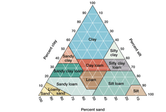
These procedure were meant to learn more about our new soil samples and get us prepared for future testing. Based on our results I can assert that my soil sample is very sandy and using the chart seen below I can assert that the soil is sand or potentially sandy loam if enough material was still in suspension. I was also assert that my soil sample was slightly acidic based on the results of my pH test. This will help my group address our question further because having a collection of soil metadata will help us determine weather or not other factors besides tree species determine phage presence.

Image result for soil pyramid(https://samanthaapes.weebly.com/apes-in-a-box-soil-pyramid.html)

Future:

This entry has been updated to reflect the results of the metadata testing; however, when the procedures stated above were completed the initial future procedures were to simply check on the results of the experiments after the appropriate amount of time had elapsed. Now that this is completed spot tests will be preformed using the lysate created using the above procedures and depending on the results of those spot tests plaque assays or more soil collection will be preformed.

September 14

Soil Washing and Metadata 9/10/2018

Rationale: wash a new soil sample to get new direct and enriched isolations. In addition, prepare the soil for analysis to get soil metadata.

Soil Washing:

  1. Filled 15 mL conical tube with ~2mL of soil
  2. Added LB the 12 mL mark of the tube
  3. Started 10 minutes of shaking and vortex-ing
    1. the tube started leaking so I tried to switch the mixture into another tube but then I switched it back when not all of the soil came out of the original tube. I ended up losing ~1mL of my LB and soil mixture
  4. After shaking I weighed the tube and got 19.83 g
  5. The tubes were centrifuged as a class
  6. I used a syringe and filter to filter the supernatant of the centrifuges mixture into a new 15 mL tube
    1. I used aseptic technique, and consistently washed the tip of the filter with 70% ethanol
    2. I lost a lot of solution to the table trying to get the last of the supernatant into the syringe
  7. I ended up getting ~7 mL for my enriched isolation and ~0.75 mL for my direct isolation, both in 15 mL conical tubes
  8. I added 0.5 mL of arthro to my enriched isolation and left it to incubate for 48 hours at 28 °C

Soil Metadata:

  1. Filled a falcon tube with ~4 mL soil
  2. Added DI water to the 12 mL mark of the tube
  3. Added 3 drops of soil dispersion
  4. shook tube with glove over the opening for 30 seconds
  5. poured supernatant into 50 mL conical tube and let both tubes sit under the hood for 48 hours
  6. measured the mass of wet soil sample in a weigh boat
    1. mass of weigh boat = 2.37g
    2. mas of wet soil = 4.62 g
  7. Took the pH of soil sample
    1. added pinch of soil to pH tube
    2. filled tube with DI
    3. shook for 10 seconds
    4. let sit for 2 min
    5. put 1 in long pH paper into tube for 45 sec
      1. dropped first paper into sample so had to use a new one
    6. pH = 6.5

Next Steps: Perform a spot test to check for phages and continue analyzing soil metadata.

September 14

9-12-18 — Spot Test and Metadata Continued

Date: Wednesday, September 12th, 2018

Title: Spot Test and Metadata Continued

Rationale: The purpose of today’s lab is to make a spot test, an agar control, and finish soil metadata.

Class Question: Is there a difference in bacteriophage presence or type in soil samples taken from live oaks vs those from red oaks?

Procedure:

  1. We began by creating an aseptic zone spreading CiDecon over the workplace then letting Ethanol (70%) evaporate off the table, dehydrating and therefore killing any organisms that could contaminate the equipment or samples.
  2. We lit a burner in the middle of the table in order to keep falling particles from contaminating the equipment or samples from above since the flame creates a circulating air current.
  3. We mixed 8 mL LB broth and 90 microliters of CaCL2 in a 50 mL conical for our plates.
  4. I used a syringe filter to transfer 1 mL of my enriched lysate to a culture tube containing .5 mL ATC 21022.
  5. Next we added 10 mL of 2X top agar to our 50 mL conical. We pipetted 4.5 mL of this solution to our top agar control plate.
  6. We quickly added 4.5 mL of our solution to our individual plates.
  7. We waited for 10 minutes for our agar to harden.
  8. We then took 10 microliters of our enriched lysate and put it on the plate. We also took 10 microliters of direct isolation and of phage buffer and added them to the plate.
  9. We let our plates sit for 15 minutes then set them in an incubator at room temperature.
  10. I took a pinch of my soil sample and added it to a small pH vial.
  11. I filled the rest of the vial with water, dipped 1 inch of pH paper in the mixture, and waited for 45 seconds.
  12. I compared the color of the paper and determined that the soil sample had a pH of 6.3.
  13. I checked my Falcon tube and determined that my sample was 75% sand, 12.5% silt, and 12.5% clay. Some silt and clay particles were still in suspension.
  14. I weighed my dry soil boat and found it to be 8.985 grams. This would make the soil by itself weigh 6.506 grams.
  15. I subtracted today’s weight from Wednesday’s 7.259 grams of wet soil and calculated that my sample was 27.6% water.

Observations:

  • My dispersion wasn’t completely settled, so soil composition percentages could be off.
  • My soil pH was 6.3, so it was slightly acidic.

Results:

  • This experiment yielded me a spot test as well as soil metadata.

Next Step:

  • My next step is to observe the results from my spot test and check for possible phage plaques.
September 14

Lab Journals for the week of September 10th

SEA Phage Lab Journal

September 10th, 2018

Metadata and Enrichment


Objectives: Collect Metadata on soil sample 2, filter lysate and separate into direct and enriched portions.


Rationale: The metadata for each tree our unit collects from can be used to better understand the way the presence of Arthrobacter phage correlates with the health of different trees. Once we have discovered correlation, we could test causation. Filtering lysate allows me to test for phage effectively.


Results from last Lab: I collected soil from a large Live Oak tree right outside of the SLC, near a very popular walkway. I have the soil sample in a 15 mL vial and a plastic bag, as well as several leaves.


Procedure: Added 2 mL of soil sample 2 into a 15 mL tube, for enrichment. The other roughly 10 mL of soil was saved in the same plastic bag it was stored in, for metadata collection. Added 10 mL of LB Broth to the 15 mL enrichment tube. Then, I shook the tube for 15 minutes to mix the soil and LB Broth together. After shaking, I massed my tube, found it to be 18.72 grams, and ended up adding several drops of DI water to match a lab partner for centrifuging at 19.22 grams.


While the tubes were in the centrifuge, I began to collect metadata. Because I didn’t collect as much soil as I should have, I couldn’t complete the proportions as instructed. Instead of filling a tube to 10 mL of soil, I filled one to 4 mL, saving roughly half of the remaining soil for later. I then added DI water to 12 mL, and 3 drops of soil dispersion liquid. Shook for 30 seconds, and let the soil settle out. Afterwards, I massed a weigh boat at 2.45 grams, and my remaining wet soil in the same boat at 8.10 grams. This means the wet soil was 5.65 grams.


The soil and LB Broth finished the 10 min in the centrifuge. I set up my filter apparatus and used a bubble pipette to move the supernatant to the filter on the top. I was only able to collect around 9 mL of lysate, so I opted to stick with just an enriched sample rather than enriched and a direct. I added .5 mL of arthrobacter and set the enriched lysate in a shaking incubator at 26 degrees Celsius for the next 48 hours.


Finally, I took a few drops of the liquid from my dispersed soil sample and added DI water until it filled a small vial. I took pH paper and tested for 45 seconds. The pH was 6.0.


Analysis and Interpretation: The soil was light colored, especially when compared with other Live Oak tree soil in the area. The pH seemed to be acidic, but I don’t know what other trees are like.


Future Plans: Next lab I will complete a spot test with my enriched lysate, and determine the need for a plaque assay the class afterwards. I will also find the percent composition of my soil, and find the proper classification based on Lathan’s chart.


SEA Phage Lab Journal

September 12th, 2018

Spot Testing and Metadata


Objectives: Complete a Spot test with my second enriched lysate, and finish soil metadata.


Rationale: Testing for plaques and a basic understanding of the environment around my tree.


Procedure: Massed enriched lysate after incubation from the last lab. Found mass buddy (21.55g), and gave the 50 mL tube to Dr. Adair to centrifuge for 5 minutes at 3,000 g. I found the mass of my dry soil in the weigh boat, now 7.38g, and found the percent water loss, at 12.7% of the original mass.


I found the dispersed soil from the last lab to be 3.5 mL inside the tube. 1.75 mL was clay, (50%),, 1 mL was silt, (28.6%), and .75 mL was sand, (21.4%). When plotted, the readout was firmly in the Clay section of the sand/silt/clay chart.


I began to filter my lysate into a microcentrifuge tube with a syringe, and a 22 micrometer filter attached to it, but as my lab partners moved around me, I drifted out of the aseptic zone and needed to repeat. I got a new syringe and filter, as well as a microcentrifuge tube. I pulled slightly less than 2 mL of enriched lysate from my 15 mL tube, attached my new filter, and pushed the plunger slowly until I had filtered everything into the microcentrifuge tube. While I was doing this, my lab partners had made an Agar plate and separated it into quadrants. We used a pipette and transferred 10 microliters of each lysate into our respective quadrants, saving one quadrant as a control. It occurred to me after lab that I didn’t notice Henry switching pipette tips, but I likely just didn’t see it. Thought it was important to note, however.


Once that was done, we let the plate sit for around 10 minutes, then set it in the incubator.


Analysis and Interpretations: I suspect my soil has a different composition from others’, due to the high amount of clay and the distinct coloration as compared to others’. I will be interested to see where other people’s soil falls on the graph.


Future Plans: Depending on how my spot test goes, I will either complete a plaque assay next lab or reevaluate my soil and determine a new place to search for samples, based on our question.